Cerveau
Un article de Vev.
(Différences entre les versions)
| Version du 3 mai 2012 à 18:44 Vev (Discuter | contribs) (→liens) ← Différence précédente |
Version du 3 mai 2012 à 18:47 Vev (Discuter | contribs) Différence suivante → |
||
| Ligne 1: | Ligne 1: | ||
| - | |||
| - | |||
| == sujet == | == sujet == | ||
| Ligne 36: | Ligne 34: | ||
| http://publishingacademy.com/wp-content/uploads/brain-mindmap.jpg | http://publishingacademy.com/wp-content/uploads/brain-mindmap.jpg | ||
| http://www.mindmapinspiration.com/wp-content/uploads/2009/02/curious-brain-mind-map-10.jpg | http://www.mindmapinspiration.com/wp-content/uploads/2009/02/curious-brain-mind-map-10.jpg | ||
| - | |||
| - | == wikipedia == | ||
| - | |||
| - | |||
| - | [[Image:Chimp Brain in a jar.jpg|thumb|Cerveau d'un [[chimpanzé]].]] | ||
| - | |||
| - | Le '''cerveau''' est le principal [[organe]] du [[système nerveux]] des [[animal|animaux]]. Au sens strict, le cerveau est l'ensemble des structures nerveuses dérivant du [[prosencéphale]] ([[diencéphale]] et [[télencéphale]]). Dans le [[langage courant]], ce terme peut désigner soit l'[[encéphale]] dans son ensemble, soit le seul [[télencéphale]] ou même le seul [[cortex cérébral]]. | ||
| - | |||
| - | Chez les [[vertébrés]], le cerveau est situé dans la [[tête (anatomie)|tête]], protégé par le [[crâne]], et son volume varie grandement d'une [[espèce (biologie)|espèce]] à l'autre. Par [[analogie]], chez les [[invertébré]]s, le cerveau désigne certains centres nerveux. | ||
| - | |||
| - | Le cerveau régule les autres [[Système d'organes|systèmes d'organes]] du corps, en agissant sur les [[muscle]]s ou les [[glande]]s, et constitue le siège des fonctions [[cognition|cognitives]]. Ce contrôle centralisé de l'organisme permet des réponses rapides et coordonnées aux variations environnementales. Les [[réflexe (réaction motrice)|réflexes]], schémas de réponses simples, ne nécessitent pas l'intervention du cerveau. Toutefois, les comportements plus sophistiqués nécessitent que le cerveau intègre les informations transmises par les [[Système sensoriel|systèmes sensoriels]] et fournissent une réponse adaptée. | ||
| - | |||
| - | Le cerveau est une structure extrêmement complexe qui peut renfermer jusqu'à plusieurs milliards de [[neurone]]s connectés les uns aux autres. Les neurones sont les [[Cellule (biologie)|cellules]] cérébrales qui communiquent entre elles par le biais de longues fibres protoplasmiques appelées [[axone]]s. L'axone d'un neurone transmet des [[influx nerveux]], les [[Potentiel d'action|potentiels d'action]], à des cellules cibles spécifiques situées dans des régions plus ou moins distantes du cerveau ou de l'organisme. Les [[cellule gliale|cellules gliales]] sont le deuxième type cellulaire du cerveau et assurent des fonctions très diversifiées, centrées autour du support des neurones et de leurs fonctions. | ||
| - | |||
| - | Malgré de grandes avancées en [[neurosciences]], le fonctionnement du cerveau est encore mal connu. Les relations qu'il entretient avec l'[[esprit]] sont le sujet de nombreuses discussions, aussi bien [[philosophique]]s que [[scientifique]]s. | ||
| - | |||
| - | == Anatomie == | ||
| - | [[Fichier:Bilaterian-plan.svg|thumb|Schéma d'organisation fondamental d'un bilatérien.]] | ||
| - | Le cerveau est la structure biologique la plus complexe connue<ref name=shepherd>{{ouvrage|langue=en|prénom1=G. M.|nom1=Shepherd|lien auteur1=|directeur1=|prénom2=|nom2=|lien auteur2=|directeur2=|prénom3=|nom3=|lien auteur3=|traducteur=|illustrateur=|préface=|titre=Neurobiology|sous-titre=|titre original=|numéro d'édition=3|collection=|série=|numéro dans collection=|lien éditeur=Oxford University Press|éditeur=Oxford University Press|lieu=|année=1994|mois=|jour=|année première édition=|réimpression=|tome=|volume=|titre volume=|pages totales=760|format=|isbn=9780195088434|isbn2=|isbn3=|issn=|issn2=|issn3=|oclc=|partie=|numéro chapitre=|titre chapitre=|passage=3|présentation en ligne=http://books.google.com/?id=zr4WRMw0xRQC|lire en ligne=|consulté le=|commentaire=|extrait=|id=|référence=|référence simplifié=}}</ref> ce qui rend souvent délicate la comparaison de cerveaux de différentes espèces à partir de leur apparence. Néanmoins, l'architecture du cerveau présente plusieurs caractéristiques communes à un grand nombre d'espèces. Trois approches complémentaires permettent de les mettre en évidence. L'approche [[Évolution (biologie)|évolutionniste]] compare l'anatomie du cerveau entre différentes espèces et repose sur le principe que les [[Caractère (biologie)|caractères]] retrouvés sur toutes les branches descendantes d'un ancêtre donné étaient aussi présentes chez leur [[ancêtre commun]]. L'approche [[Neurodéveloppement|développementale]] étudie le processus de formation du cerveau du stade [[embryon]]naire au stade [[adulte]]. Enfin, l'approche [[génétique]] analyse l'[[Expression génétique|expression]] des [[gène]]s dans les différentes zones du cerveau. | ||
| - | |||
| - | L'origine du cerveau remonte à l'apparition des [[Bilateria|bilatériens]], une des principales subdivisions du [[règne animal]] notamment caractérisée par une [[symétrie bilatérale]] des organismes, il y a environ 550-560 [[Million d'années|millions d'années]]<ref>{{article|langue=en|prénom1=G.|nom1=Balavoine|lien auteur1=|prénom2=A.|nom2=Adoutte|lien auteur2=|prénom3=|nom3=|lien auteur3=|traduction=|titre=The Segmented Urbilateria: A Testable Scenario|sous-titre=|périodique=Integr. Comp. Biol.|lien périodique=Integrative and Comparative Biology|éditeur=|lieu=|série=|volume=43|titre volume=|numéro=1|titre numéro=|jour=|mois=|année=2003|pages=137-147|issn=1540-7063|issn2=1557-7023|issn3=|isbn=|résumé=|format=|url texte=http://icb.oxfordjournals.org/content/43/1/137.full.pdf+html|doi=10.1093/icb/43.1.137| consulté le=|commentaire=|extrait=|id=}}</ref>. L'ancêtre commun de ce taxon suivait un [[plan d'organisation]] de type [[Tube|tubulaire]], [[ver]]miforme et [[Métamérisation|métamérisé]] ; un schéma qui continue de se retrouver dans le corps de tous les bilatériens actuels, dont l'[[Homme]]<ref>{{ouvrage|langue=en|prénom1=A.|nom1=Schmidt-Rhaesa|lien auteur1=|directeur1=|prénom2=|nom2=|lien auteur2=|directeur2=|prénom3=|nom3=|lien auteur3=|traducteur=|illustrateur=|préface=|titre=The evolution of organ systems|sous-titre=|titre original=|numéro d'édition=|collection=|série=|numéro dans collection=|lien éditeur=Oxford University Press|éditeur=Oxford University Press|lieu=|année=2007|mois=|jour=|année première édition=|réimpression=|tome=|volume=|titre volume=|pages totales=385|format=|isbn=9780198566694|isbn2=|isbn3=|issn=|issn2=|issn3=|oclc=|partie=|numéro chapitre=|titre chapitre=|passage=110|présentation en ligne=http://books.google.com/books?id=ZACR7ZO_65YC&hl=fr|lire en ligne=|consulté le=|commentaire=|extrait=|id=|référence=|référence simplifié=}}</ref>. Ce plan d'organisation fondamental du corps est un [[tube]] renfermant un [[tube digestif]], reliant la [[bouche]] et l'[[anus]], et un [[cordon nerveux]] qui porte un [[Ganglion nerveux|ganglion]] au niveau de chaque métamère du corps et notamment un ganglion plus important au niveau du front appelé « cerveau ». | ||
| - | |||
| - | === Invertébrés === | ||
| - | |||
| - | La composition du cerveau des [[invertébrés]] est très différente de celle des [[vertébrés]], à tel point qu'il est difficile de comparer les deux structures à moins de se baser sur la [[génétique]]. Deux groupes d'invertébrés se démarquent par un cerveau relativement complexe : les [[arthropodes]] et les [[céphalopodes]]<ref name="butler">{{article|langue=en|prénom1=A. B.|nom1=Butler|lien auteur1=|prénom2=|nom2=|lien auteur2=|prénom3=|nom3=|lien auteur3=|traduction=|titre=Chordate evolution and the origin of craniates: An old brain in a new head|sous-titre=|périodique=Anat. Rec.|lien périodique=Anatomical Record|éditeur=|lieu=|série=|volume=261|titre volume=|numéro=3|titre numéro=|jour=15|mois=juin|année=2000|pages=111-125|issn=1932-8494|issn2=|issn3=|isbn=|résumé=|format=|url texte=|doi=10.1002/1097-0185(20000615)261:3<111::AID-AR6>3.0.CO;2-F| consulté le=|commentaire=|extrait=|id=}}</ref>. Le cerveau de ces deux groupes provient de deux cordons nerveux parallèles qui s'étendent à travers tout le corps de l'animal. Les arthropodes ont un cerveau central avec trois divisions et de larges lobes optiques derrière chaque [[œil]] pour le [[Vue|traitement visuel]]<ref name="butler"/>. Les céphalopodes possèdent le plus gros cerveau de tous les invertébrés. Le cerveau des pieuvres est très développé, avec une complexité similaire à celle rencontrée chez les vertébrés. | ||
| - | |||
| - | Le cerveau de quelques invertébrés a été particulièrement étudié. Par la simplicité et l'accessibilité de son système nerveux, l'[[aplysie]] a été choisie comme modèle par le [[Neurophysiologie|neurophysiologiste]] [[Eric Kandel]] pour l'étude des bases moléculaires de la mémoire qui lui valut un [[Prix Nobel de physiologie ou médecine|Prix Nobel]] en [[2000]]<ref>{{ouvrage|langue=en|prénom1=E. R.|nom1=Kandel|lien auteur1=|directeur1=|prénom2=|nom2=|lien auteur2=|directeur2=|prénom3=|nom3=|lien auteur3=|traducteur=|illustrateur=|préface=|titre=In search of memory: the emergence of a new science of mind|sous-titre=|titre original=|numéro d'édition=|collection=|série=|numéro dans collection=|lien éditeur=|éditeur=W. W. Norton & Co.|lieu=|année=2007|mois=|jour=|année première édition=|réimpression=|tome=|volume=|titre volume=|pages totales=510|format=|isbn=9780393329377|isbn2=|isbn3=|issn=|issn2=|issn3=|oclc=|partie=|numéro chapitre=|titre chapitre=|passage=|présentation en ligne=http://books.google.com/books?id=LURy5gojaDoC&hl=fr|lire en ligne=|consulté le=|commentaire=|extrait=|id=|référence=|référence simplifié=}}</ref>. Cependant, les cerveaux d'invertébrés les plus étudiés demeurent ceux de la [[drosophile]] et du [[Nematoda|ver nématode]] ''[[Caenorhabditis elegans]]''. Du fait de l'important panel de techniques à disposition pour étudier leur [[matériel génétique]], les drosophiles sont tout naturellement devenues un sujet d'étude sur le rôle des [[gène]]s dans le développement du cerveau<ref>{{Lien web|url=http://flybrain.neurobio.arizona.edu/ |titre=Flybrain: An online atlas and database of the ''drosophila'' nervous system|id= |série= |auteur= |lien auteur= |coauteurs= |date= |année= |mois= |site= |éditeur=Département d'entomologie de l'université du Colorado |isbn= |page= |citation= |en ligne le= |consulté le=29 octobre 2010 }}</ref>. De nombreux aspects de la neurogénétique des drosophiles se sont avéré être également valable chez l'Homme. Par exemple, les premiers gènes impliqués dans l'[[horloge circadienne|horloge biologique]] furent identifiés dans les [[années 1970]] en étudiant des drosophiles mutantes montrant des perturbations dans leur cycles journaliers d'activité<ref>{{article|langue=en|prénom1=R. J.|nom1=Konopka|lien auteur1=|prénom2=S.|nom2=Benzer|lien auteur2=|prénom3=|nom3=|lien auteur3=|traduction=|titre=Clock Mutants of ''Drosophila melanogaster''|sous-titre=|périodique=Proc. Natl. Acad. Sci. USA|lien périodique=Proceedings of the National Academy of Sciences|éditeur=|lieu=|série=|volume=68|titre volume=|numéro=9|titre numéro=|jour=1|mois=septembre|année=1971|pages=2112-2116|issn=0027-8424|issn2=|issn3=|isbn=|résumé=|format=|url texte=http://www.pnas.org/content/68/9/2112.full.pdf+html|doi=| consulté le=|commentaire=|extrait=|id=}}</ref>. Une recherche sur le [[génome]] des vertébrés a montré un ensemble de [[Analogie (évolution)|gènes analogues]] à ceux de la drosophile jouant un rôle similaire dans l'horloge biologique de la souris et probablement également dans celle de l'Homme<ref>{{article|langue=en|prénom1=H.-S.|nom1=Shin|lien auteur1=|prénom2=T. A.|nom2=Bargiello|lien auteur2=|prénom3=B. T.|nom3=Clark|lien auteur3=|prénom4=F. R.|nom4=Jackson|lien auteur4=|prénom5=M. W.|nom5=Young|lien auteur5=|traduction=|titre=An unusual coding sequence from a ''Drosophila'' clock gene is conserved in vertebrates|sous-titre=|périodique=Nature|lien périodique=Nature (revue)|éditeur=|lieu=|série=|volume=317|titre volume=|numéro=6036|titre numéro=|jour=3|mois=octobre|année=1985|pages= 445-448|issn=0028-0836|issn2=|issn3=|isbn=|résumé=|format=|url texte=|doi=10.1038/317445a0| consulté le=|commentaire=|extrait=|id=}}</ref>. | ||
| - | Comme la drosophile, le nématode ''C. elegans'' a été très étudié en génétique<ref>{{Lien web|url= http://www.wormbook.org/|titre=WormBook: The online review of ''C. Elegans'' biology |id= |série= |auteur= |lien auteur= |coauteurs= |date= |année= |mois= |site= |éditeur= |isbn= |page= |citation= |en ligne le= |consulté le= 30 octobre 2010}}</ref> car son plan d'organisation est très stéréotypé : le système nerveux du morphe [[Hermaphrodisme|hermaphrodite]] possède exactement 302 [[neurone]]s, toujours à la même place, établissant les mêmes [[Synapse|liaisons synaptiques]] pour chaque ver<ref>{{article|langue=en|prénom1=O.|nom1=Hobert|lien auteur1=|prénom2=|nom2=|lien auteur2=|prénom3=|nom3=|lien auteur3=|traduction=|titre=Specification of the nervous system|sous-titre=|périodique=WormBook|lien périodique=WormBook|éditeur=The C. elegans Research Community|lieu=|série=|volume=|titre volume=|numéro=|titre numéro=|jour=8|mois=août|année=2005|pages=1-19|issn=|issn2=|issn3=|isbn=|résumé=|format=|url texte=http://www.wormbook.org/chapters/www_specnervsys/specnervsys.html|doi=10.1895/wormbook.1.12.1| consulté le=26 décembre 2010|commentaire=|extrait=|id=}}</ref>. Au début des années 1970, du fait de sa simplicité et de sa facilité d’élevage, [[Sydney Brenner]] le choisit comme [[organisme modèle]] pour ses travaux sur le processus de régulation génétique du développement qui lui valurent un Prix Nobel en [[2002]]<ref>{{en}} {{Lien web|url=http://elegans.swmed.edu/Sydney.html |titre=Sydney Brenner |id= |série= |auteur= |lien auteur= |coauteurs= |date= |année=1987 |mois= |site= |éditeur= |isbn= |page= |citation= |en ligne le= |consulté le=26 décembre 2010 }}</ref>. Pour ses travaux, Brenner et son équipe ont découpé les vers en milliers de sections ultra fines et photographié chacune d'entre elles au [[Microscopie électronique|microscope électronique]] afin de visualiser les fibres assorties à chaque section et ainsi planifier chaque neurone et chaque synapse dans le corps du ver<ref>{{article|langue=en|prénom1=J. G.|nom1=White|lien auteur1=|prénom2=E.|nom2=Southgate|lien auteur2=|prénom3=J. N.|nom3=Thomson|lien auteur3=|prénom4=S.|nom4=Brenner|lien auteur4=Sydney Brenner|traduction=|titre=The Structure of the Nervous System of the Nematode Caenorhabditis elegans|sous-titre=|périodique=Phil. Trans. R. Soc. B|lien périodique=Phil. Trans. R. Soc. B|éditeur=|lieu=|série=|volume=314|titre volume=|numéro=1165|titre numéro=|jour=12|mois=novembre|année=1986|pages=1-340|issn=1471-2970|issn2=|issn3=|isbn=|résumé=|format=|url texte=|doi=10.1098/rstb.1986.0056| consulté le=|commentaire=|extrait=|id=}}</ref>. Actuellement, un tel niveau de détail n'est disponible pour aucun autre organisme, et les informations récoltées ont rendu possibles de nombreuses études. | ||
| - | |||
| - | === Vertébrés === | ||
| - | [[Image:Brains-fr.svg|thumb|Comparaison des cerveaux de différentes espèces]] | ||
| - | Apparus il y a 500 millions d'années, les [[vertébrés]] ont dérivé d'une forme proche de la [[myxine]] actuelle<ref>{{article|langue=en|prénom1=D.-G.|nom1=Shu|lien auteur1=|prénom2=S.|nom2=Conway Morris|lien auteur2=|prénom3=J.|nom3=Han|lien auteur3=|prénom4=Z.-F.|nom4=Zhang|lien auteur4=|prénom5=K.|nom5=Yasui|lien auteur5=|prénom6=P.|nom6=Janvier|lien auteur6=|prénom7=L.|nom7=Chen|lien auteur7=|prénom8=X.-L.|nom8=Zhang|lien auteur8=|prénom9=J.-N|nom9=Liu|lien auteur9=|prénom10=Y.|nom10=Li|lien auteur10=|prénom11=H.-Q|nom11=Liu|lien auteur11=|traduction=|titre=Head and backbone of the Early Cambrian vertebrate Haikouichthys|sous-titre=|périodique=Nature|lien périodique=Nature (revue)|éditeur=|lieu=|série=|volume=421|titre volume=|numéro=6922|titre numéro=|jour=30|mois=janvier|année=2003|pages=526-529|issn=0028-0836|issn2=|issn3=|isbn=|résumé=|format=|url texte=|doi=10.1038/nature01264| consulté le=|commentaire=|extrait=|id=}}</ref>. Le cerveau de tous les vertébrés présente fondamentalement la même structure<ref>{{ouvrage|langue=fr|prénom1=R.|nom1=Eckert|lien auteur1=|directeur1=|prénom2=D.|nom2=Randall|lien auteur2=|directeur2=|prénom3=|nom3=|lien auteur3=|traducteur=F. Math|illustrateur=|préface=|titre=Physiologie animale|sous-titre=Mécanismes et adaptations|titre original=|numéro d'édition=4|collection=|série=|numéro dans collection=|lien éditeur=De Boeck Université|éditeur=De Boeck Université|lieu=|année=1999|mois=|jour=|année première édition=|réimpression=|tome=|volume=|titre volume=|pages totales=822|format=|isbn=9782744500534|isbn2=|isbn3=|issn=|issn2=|issn3=|oclc=|partie=|numéro chapitre=|titre chapitre=|passage=415|présentation en ligne=http://books.google.fr/books?id=OaSHXG2liFgC&dq=Physiologie+animale+:+m%C3%A9canismes+et+adaptations&source=gbs_navlinks_s|lire en ligne=http://books.google.fr/books?id=OaSHXG2liFgC&lpg=PP1&dq=Physiologie%20animale%20%3A%20m%C3%A9canismes%20et%20adaptations&pg=PA415#v=onepage&q&f=false|consulté le={{1er}} janvier 2011|commentaire=|extrait=|id=|référence=|référence simplifié=}}</ref>. Au cours de l'évolution de cet embranchement, la tendance évolutive du cerveau a été de suivre un gradient de taille et de complexité croissante, en particulier chez les [[mammifères]]<ref>{{ouvrage|langue=en|prénom1=G. F.|nom1=Striedter|lien auteur1=|directeur1=|prénom2=|nom2=|lien auteur2=|directeur2=|prénom3=|nom3=|lien auteur3=|traducteur=|illustrateur=|préface=|titre=Principles of brain evolution|sous-titre=|titre original=|numéro d'édition=|collection=|série=|numéro dans collection=|lien éditeur=|éditeur=Sinauer Associates|lieu=|année=2005|mois=|jour=|année première édition=|réimpression=|tome=|volume=|titre volume=|pages totales=436|format=|isbn=9780878938209|isbn2=|isbn3=|issn=|issn2=|issn3=|oclc=|partie=|numéro chapitre=|titre chapitre=|passage=|présentation en ligne=http://books.google.fr/books?id=JzdmQgAACAAJ&dq=isbn:9780878938209&ei=ET4fTYLeDoOsywSA4JXrCw&cd=1|lire en ligne=|consulté le=|commentaire=|extrait=|id=|référence=|référence simplifié=}}</ref>. | ||
| - | |||
| - | Le cerveau des vertébrés est d'un [[tissu mou]] et d'une texture [[gélatine]]use<ref>{{ouvrage|langue=en|prénom1=E. R.|nom1=Kandel|lien auteur1=|directeur1=|prénom2=J. H.|nom2=Schwartz|lien auteur2=|directeur2=|prénom3=T. M.|nom3=Jessell|lien auteur3=|traducteur=|illustrateur=|préface=|titre=Principles of neural science|sous-titre=|titre original=|numéro d'édition=4|collection=|série=|numéro dans collection=|lien éditeur=McGraw-Hill|éditeur=McGraw-Hill|lieu=|année=2000|mois=|jour=|année première édition=|réimpression=|tome=|volume=|titre volume=|pages totales=1414|format=|isbn=9780838577011|isbn2=|isbn3=|issn=|issn2=|issn3=|oclc=|partie=|numéro chapitre=17|titre chapitre=|passage=|présentation en ligne=http://books.google.fr/books?id=yMtpAAAAMAAJ&dq=isbn:9780838577011&ei=-j4fTejhBNPaUM_hzZYI&cd=1|lire en ligne=|consulté le=|commentaire=|extrait=|id=Kandel|référence=|référence simplifié=}}</ref>. De manière générale, le tissu cérébral vivant est rosâtre à l'extérieur et blanchâtre à l'intérieur. Le cerveau des vertébrés est enveloppé d'un [[Membrane (biologie)|système membranaire]] de [[tissu conjonctif]], les [[méninge]]s, qui sépare le [[crâne]] du cerveau<ref>{{ouvrage|langue=en|prénom1=A.|nom1=Parent|lien auteur1=|directeur1=|prénom2=M. B.|nom2=Carpenter|lien auteur2=|directeur2=|prénom3=|nom3=|lien auteur3=|traducteur=|illustrateur=|préface=|titre=Carpenter's human neuroanatomy|sous-titre=|titre original=|numéro d'édition=9|collection=|série=|numéro dans collection=|lien éditeur=|éditeur=Williams & Wilkins|lieu=|année=1996|mois=|jour=|année première édition=|réimpression=|tome=|volume=|titre volume=|pages totales=1011|format=|isbn=9780683067521|isbn2=|isbn3=|issn=|issn2=|issn3=|oclc=|partie=|numéro chapitre=1|titre chapitre=|passage=|présentation en ligne=http://books.google.com/books?id=IJ5pAAAAMAAJ&hl=fr|lire en ligne=|consulté le=|commentaire=|extrait=|id=|référence=|référence simplifié=}}</ref>. De l'extérieur vers l'intérieur, les méninges sont composées de trois membranes : la [[dure-mère]], l'[[arachnoïde]] et la [[pie-mère]]. L'arachnoïde et la pie-mère sont étroitement connectées entre elles et peuvent ainsi être considérées comme une seule et même couche, la pie-arachnoïde. Compris entre l'arachnoïde et la pie mère, l'espace sous-arachnoïdien contient le [[liquide cérébro-spinal]] qui circule dans l'étroit espace entre les cellules et à travers les cavités appelées [[système ventriculaire]]. Ce liquide sert notamment de protection mécanique au cerveau en absorbant et amortissant les chocs et à transporter [[hormone]]s et [[nutriment]]s vers le tissu cérébral. Les [[Vaisseau sanguin|vaisseaux sanguins]] viennent irriguer le [[système nerveux central]] à travers l'espace périvasculaire au-dessus de la pie-mère. Au niveau des vaisseaux sanguins, les cellules sont étroitement jointes, formant la [[barrière hémato-encéphalique]] qui protège le cerveau en agissant comme un [[filtre]] vis-à-vis des [[toxine]]s susceptibles d'être contenues dans le [[sang]]. | ||
| - | |||
| - | Tous les cerveaux des vertébrés possèdent la même forme sous-jacente observable par la manière dont le cerveau se développe<ref>{{Modèle:Référence Harvard sans parenthèses | Kandel | 2000 | p=1019 | id=Kandel}}</ref>. Pendant le [[neurodéveloppement]], le système nerveux commence à se mettre en place par l'apparition d'une fine bande de tissu neural parcourant tout le [[dos]] de l'[[embryon]]. La bande s'épaissit ensuite et se plisse pour former le [[tube neural]]. C'est à l'extrémité crânienne du tube que se développe le cerveau, l'émergence de ce crâne chez les premiers vertébrés aquatiques étant en relation avec le développement de leur sens de l'[[olfaction]] lié à leur capacités exploratrices à la recherche de proies. Au départ, le cerveau se manifeste comme trois gonflements qui représentent en fait le [[prosencéphale]], le [[mésencéphale]] et le [[rhombencéphale]]. Chez de nombreux groupes de vertébrés, ces trois régions gardent la même taille chez l'adulte, mais le prosencéphale des mammifères devient plus important que les autres régions et le mésencéphale plus petit<ref name="Vincent">{{ouvrage|auteur=[[Jean-Didier Vincent]] Pierre-Marie Liedo|titre=Le cerveau sur mesure|éditeur=Odile Jacob|date=2012|pages totales=290|isbn=2738127096|lire en ligne=}}</ref>. | ||
| - | |||
| - | La relation entre la taille du cerveau, la taille de l'organisme et d'autres facteurs a été étudiée à travers un grand nombre d'espèces de vertébrés. La taille du cerveau augmente avec la taille de l'organisme, mais pas de manière proportionnelle. Chez les mammifères, la relation suit une [[loi de puissance]], avec un [[Exposant (mathématiques)|exposant]] d'environ 0,75<ref>{{article|langue=en|prénom1=E.|nom1=Armstrong|lien auteur1=|prénom2=|nom2=|lien auteur2=|prénom3=|nom3=|lien auteur3=|traduction= |titre= | ||
| - | Relative Brain Size and Metabolism in Mammals | ||
| - | |sous-titre=|périodique=Science|lien périodique=Science (revue)|éditeur=|lieu=|série=|volume=220|titre volume=|numéro=4603|titre numéro=|jour=17|mois=juin|année=1983|pages=1302-1304|issn=0036-8075|issn2=|issn3=|isbn=|résumé=|format=|url texte=|doi=10.1126/science.6407108| consulté le=|commentaire=|extrait=|id=}}</ref>. Cette formule s'applique pour le cerveau moyen des mammifères mais chaque famille s'en démarque plus ou moins, reflétant la complexité de leur comportement. Ainsi, les [[primates]] ont un cerveau cinq à dix fois plus gros que ce qu’indique la formule. De manière générale, les [[Prédation|prédateurs]] tendent à avoir des cerveaux plus gros. Quand le cerveau des mammifères augmente en taille, toutes les parties n'augmentent pas dans la même proportion. Plus le cerveau d'une espèce est gros, plus la fraction occupée par le [[cortex]] est importante<ref name="finlay">{{article|langue=en|prénom1=B. L.|nom1=Finlay|lien auteur1=|prénom2=R. B.|nom2=Darlington|lien auteur2=|prénom3=N.|nom3=Nicastro|lien auteur3=|traduction=|titre=Developmental structure in brain evolution|sous-titre=|périodique=Behav. Brain Sci.|lien périodique=Behavioral and Brain Sciences|éditeur=|lieu=|série=|volume=24|titre volume=|numéro=2|titre numéro=|jour=|mois=|année=2001|pages=263-278|issn=|issn2=|issn3=|isbn=|résumé=|format=|url texte=|doi=10.1017/S0140525X01003958| consulté le=|commentaire=|extrait=|id=}}</ref>, 80% de l'activité cérébrale dépendant des signaux visuels chez les primates<ref name="Vincent"/>. | ||
| - | |||
| - | ==== Régions du cerveau ==== | ||
| - | |||
| - | En [[neuroanatomie]] des vertébrés, le cerveau est généralement considéré comme constitué de six régions principales définies sur la base du développement du système nerveux à partir du [[tube neural]] : le [[télencéphale]], le [[diencéphale]], le [[mésencéphale]], le [[cervelet]], le [[Pont (système nerveux)|pont]], et le [[bulbe rachidien]]<ref>{{Modèle:Référence Harvard sans parenthèses | Kandel | 2000 | loc=ch. 17 | id=Kandel}}</ref>. Chacune de ces régions possède une structure interne complexe. Certaines régions du cerveau, comme le [[cortex cérébral]] ou le cervelet, sont formés de couches formant des replis sinueux, les [[Circonvolution cérébrale|circonvolutions cérébrales]], qui permettent d'augmenter la surface corticale tout en logeant dans la boîte crânienne. Les autres régions du cerveau représentent des groupes de nombreux [[Noyau (biologie)|noyaux]]. Si des distinctions claires peuvent être établies à partir de la structure neurale, la chimie et la connectivité, des milliers de régions distinctes peuvent être identifiées dans le cerveau des vertébrés. | ||
| - | |||
| - | Chez plusieurs branches des vertébrés, l'évolution a amené des changements importants sur l'architecture du cerveau. Les composants du cerveau des [[requin]]s sont assemblés de façon simple et directe, mais chez les [[Téléostéens|poissons téléostéens]], groupe majoritaire des poissons modernes, le [[prosencéphale]] est devenu [[wikt:éverté|éverté]]. Le cerveau des oiseaux présente également d'importants changements<ref>{{article|langue=en|prénom1=R. G.|nom1=Northcutt|lien auteur1=|prénom2=|nom2=|lien auteur2=|prénom3=|nom3=|lien auteur3=|traduction=|titre=Forebrain evolution in bony fishes|sous-titre=|périodique=Brain Res. Bull.|lien périodique=Brain Research Bulletin|éditeur=|lieu=|série=|volume=75|titre volume=|numéro=2-4|titre numéro=|jour=18|mois=mars|année=2008|pages=191-205|issn=0361-9230|issn2=|issn3=|isbn=|résumé=|format=|url texte=|doi=10.1016/j.brainresbull.2007.10.058| consulté le=|commentaire=|extrait=|id=}}</ref>. Un des principaux composants du prosencéphale des oiseaux, la crête ventriculaire dorsale, a longtemps été considéré comme l'équivalent du [[Ganglions de la base|ganglion basal]] des mammifères, mais est maintenant considéré comme étroitement apparenté au [[néocortex]]<ref>{{article|langue=en|prénom1=A.|nom1=Reiner|lien auteur1=|prénom2=K.|nom2=Yamamoto|lien auteur2=|prénom3=H. J.|nom3=Karten|lien auteur3=|traduction=|titre=Organization and evolution of the avian forebrain|sous-titre=|périodique=Anat. Rec.|lien périodique=Anat. Rec.|éditeur=|lieu=|série=|volume=287A|titre volume=|numéro=1|titre numéro=|jour=|mois=novembre|année=2005|pages=1080–1102|issn=1932-8494|issn2=|issn3=|isbn=|résumé=|format=|url texte=|doi=10.1002/ar.a.20253|pmid=16206213| consulté le=|commentaire=|extrait=|id=}}</ref>. | ||
| - | |||
| - | De nombreuses régions du cerveau ont gardé les mêmes propriétés chez tous les vertébrés<ref name=shepherd />. Le rôle de la plupart de ces régions est encore soumis à la discussion mais il est malgré tout possible de dresser une liste des régions principales du cerveau et le rôle qu'on leur attribue selon les connaissances actuelles : | ||
| - | |||
| - | [[Image:Vertebrate-brain-regions.png|vignette|Les principales divisions de l'encéphale représentées sur un cerveau de [[requin]] et un cerveau humain]] | ||
| - | |||
| - | * Le [[bulbe rachidien]] (ou ''medulla oblongata'') prolonge la [[moelle épinière]]. Elle contient de nombreux petits [[Noyau (biologie)|noyaux]] impliqués dans un grand nombre de fonctions [[Mouvement (anatomie)|motrices]] et [[Sens (physiologie)|sensitives]]<ref>{{Modèle:Référence Harvard sans parenthèses | Kandel | 2000 | loc=ch. 44-45 | id=Kandel}}</ref>. | ||
| - | * L'[[hypothalamus]] est un petit [[organe]] situé sous le [[prosencéphale]]. Il est composé de nombreux petits noyaux possédant chacun ses propres connexions et une [[neurochimie]] particulière. L'hypothalamus régule et contrôle de nombreuses fonctions biologiques essentielles telles que l'[[Sommeil|éveil]] et le [[sommeil]], la [[faim]] et la [[soif]], ou la libération d'[[hormone]]s<ref>{{ouvrage|langue=en|prénom1=D. F.|nom1=Swaab|lien auteur1=|directeur1=|prénom2=|nom2=|lien auteur2=|directeur2=|prénom3=|nom3=|lien auteur3=|traducteur=|illustrateur=|préface=|titre=The human hypothalamus|sous-titre=Handbook of clinical neurology. Neuropathology of the human hypothalamus and adjacent brain structure|titre original=|numéro d'édition=|collection=|série=|numéro dans collection=|lien éditeur=Elsevier Science|éditeur=Elsevier Health Sciences|lieu=|année=2004|mois=|jour=|année première édition=|réimpression=|tome=|volume=2|titre volume=|pages totales=597|format=|isbn=9780444514905|isbn2=|isbn3=|issn=|issn2=|issn3=|oclc=|partie=|numéro chapitre=|titre chapitre=|passage=|présentation en ligne=http://books.google.com/books?id=Js81Pr1PmaAC&hl=fr|lire en ligne=|consulté le=|commentaire=|extrait=|id=|référence=|référence simplifié=}}</ref>. | ||
| - | * Le [[thalamus]] est également composé de noyaux aux fonctions diverses. Une partie d'entre eux servent à relayer l'information entre les [[Hémisphère cérébral|hémisphères cérébraux]] et le [[tronc cérébral]]. D'autres sont impliqués dans la [[motivation]]. La ''[[zona incerta]]'', ou région sous-thalamique, semble jouer un rôle dans plusieurs comportements élémentaires comme la [[faim]], la [[soif]], la [[défécation]] et la [[copulation]]<ref>{{ouvrage|langue=en|prénom1=E. G.|nom1=Jones|lien auteur1=|directeur1=|prénom2=|nom2=|lien auteur2=|directeur2=|prénom3=|nom3=|lien auteur3=|traducteur=|illustrateur=|préface=|titre=The thalamus|sous-titre=|titre original=|numéro d'édition=|collection=|série=|numéro dans collection=|lien éditeur=Springer Verlag|éditeur=Plenum Press|lieu=|année=1985|mois=|jour=|année première édition=|réimpression=|tome=|volume=|titre volume=|pages totales=935|format=|isbn=9780306418563|isbn2=|isbn3=|issn=|issn2=|issn3=|oclc=|partie=|numéro chapitre=|titre chapitre=|passage=|présentation en ligne=http://books.google.com/books?id=WMxqAAAAMAAJ&hl=fr|lire en ligne=|consulté le=|commentaire=|extrait=|id=|référence=|référence simplifié=}}</ref>. | ||
| - | * Le [[cervelet]] joue un rôle majeur dans la coordination des mouvements en modulant et optimisant les informations provenant d'autres régions cérébrales afin de les rendre plus précises. Cette précision n'est pas acquise à la naissance et s'apprend avec l'[[expérience]]<ref>{{Modèle:Référence Harvard sans parenthèses | Kandel | 2000 | loc=ch. 42 | id=Kandel}}</ref>. | ||
| - | * Le [[tectum]], partie supérieure du [[mésencéphale]], permet de diriger les actions dans l'espace et de conduire le mouvement. Chez les mammifères, l'aire du tectum la plus étudiée est le [[colliculus supérieur]] qui s'occupe de diriger le mouvement des [[yeux]]. Le tectum reçoit de nombreuses informations visuelles, mais aussi les informations d'autres sens qui peuvent être utiles pour diriger les actions comme l'[[ouïe]]. Chez certains poissons, comme la [[lamproie]], le tectum occupe la plus large partie du cerveau<ref>{{article|langue=en|prénom1=K.|nom1=Saitoh|lien auteur1=|prénom2=A.|nom2=Ménard|lien auteur2=|prénom3=S.|nom3=Grillner|lien auteur3=|traduction=|titre=Tectal control of locomotion, steering, and eye movements in lamprey|sous-titre=|périodique=J. Neurophysiol.|lien périodique=Journal of Neurophysiology|éditeur=|lieu=|série=|volume=97|titre volume=|numéro=4|titre numéro=|jour=|mois=avril|année=2007|pages=3093-3108|issn=0022-3077|issn2=|issn3=|isbn=|résumé=|format=|url texte=http://jn.physiology.org/content/97/4/3093.full.pdf|pmid = 17303814|doi=10.1152/jn.00639| consulté le=|commentaire=|extrait=|id=}}</ref>. | ||
| - | * Le [[Pallium (anatomie)|pallium]] est une couche de [[Substance grise|matière grise]] qui s'étale sur la surface du [[prosencéphale]]. Chez les mammifères et les reptiles, il est appelé [[cortex cérébral]]. Le pallium est impliqué dans de nombreuses fonctions telles que l'[[olfaction]] et la [[Mémoire (psychologie)|mémoire spatiale]]. Chez les mammifères, il s'agit de la région dominante du cerveau et elle [[wikt:subsumer|subsume]] les fonctions de nombreuses [[Sous-cortical|régions sous-corticales]]<ref>{{article|langue=en|prénom1=L.|nom1=Puelles|lien auteur1=|prénom2=|nom2=|lien auteur2=|prénom3=|nom3=|lien auteur3=|traduction=|titre=Thoughts on the development, structure and evolution of the mammalian and avian telencephalic pallium|sous-titre=|périodique=Phil. Trans. R. Soc. B|lien périodique=Phil. Trans. R. Soc. B|éditeur=|lieu=|série=|volume=356|titre volume=|numéro=1414|titre numéro=|jour=29|mois=octobre|année=2001|pages=1583-1598|issn=0962-8436|issn2=|issn3=|isbn=|résumé=|format=|url texte=http://rstb.royalsocietypublishing.org/content/356/1414/1583.full.pdf|pmid=11604125 |doi=10.1098/rstb.2001.0973| consulté le=|commentaire=|extrait=|id=}}</ref>. | ||
| - | * L'[[Hippocampe (cerveau)|hippocampe]], au sens strict, n'est présent que chez les mammifères. Néanmoins, cette région dérive du pallium médial communs à tous les vertébrés. Sa fonction est encore mal connue mais cette partie du cerveau intervient dans la mémoire spatiale et la navigation<ref>{{article|langue=en|prénom1=C.|nom1=Salas|lien auteur1=|prénom2=C.|nom2=Broglio|lien auteur2=|prénom3=F.|nom3=Rodríguez|lien auteur3=|traduction=|titre=Evolution of Forebrain and Spatial Cognition in Vertebrates: Conservation across Diversity|sous-titre=|périodique=Brain Behav. Evol.|lien périodique=Brain Behavior and Evolution|éditeur=|lieu=|série=|volume=62|titre volume=|numéro=2|titre numéro=|jour=|mois=|année=2003|pages=72-82|issn= 0006-8977|issn2=1421-9743|issn3=|isbn=|résumé=|format=|url texte=|doi = 10.1159/000072438| pmid = 12937346| consulté le=|commentaire=|extrait=|id=}}</ref>. | ||
| - | * Les [[ganglions de la base]] sont un groupe de structures interconnectées situées dans le prosencéphale. La fonction principale de ces ganglions semble être la [[sélection de l'action]]. Ils envoient des signaux inhibiteurs à toutes les parties du cerveau qui peuvent générer des actions et, dans les bonnes circonstances, peuvent lever l'inhibition afin de débloquer le processus et permettre l'exécution de l'action. Les [[Système de récompense|récompenses]] et les [[punition]]s exercent leurs plus importants effets neuraux au niveau des ganglions de la base<ref>{{article|langue=|prénom1=S.|nom1=Grillner|lien auteur1=|prénom2=J.|nom2=Hellgren|lien auteur2=|prénom3=A.|nom3=Ménard|lien auteur3=|prénom4=K.|nom4=Saitoh|lien auteur4=|prénom5=M. A.|nom5=Wikström|lien auteur5=|traduction=|titre=Mechanisms for selection of basic motor programs – roles for the striatum and pallidum|sous-titre=|périodique=Trends Neurosci.|lien périodique=Trends in Neurosciences|éditeur=|lieu=|série=|volume=28|titre volume=|numéro=7|titre numéro=|jour=|mois=juillet|année=2005|pages=364-370|issn=0166-2236|issn2=|issn3=|isbn=|résumé=|format=|url texte=|pmid = 15935487|doi=10.1016/j.tins.2005.05.004 | consulté le=|commentaire=|extrait=|id=}}</ref>. | ||
| - | * Le [[bulbe olfactif]] est une structure particulière qui traite les signaux olfactifs et envoie l'information vers la zone olfactive du pallium. Chez beaucoup de vertébrés, le bulbe olfactif est très développé mais il est plutôt réduit chez les [[Primates]]<ref>{{article|langue=en|prénom1=R. G.|nom1=Northcutt|lien auteur1=|prénom2=|nom2=|lien auteur2=|prénom3=|nom3=|lien auteur3=|traduction=|titre=Evolution of the Telencephalon in Nonmammals|sous-titre=|périodique=Ann. Rev. Neurosci.|lien périodique=Annual Review of Neuroscience|éditeur=|lieu=|série=|volume=4|titre volume=|numéro=|titre numéro=|jour=|mois=mars|année=1981|pages=301-350|issn= 0147-006X|issn2=|issn3=|isbn=|résumé=|format=|url texte=|pmid=7013637|doi=10.1146/annurev.ne.04.030181.001505| consulté le=|commentaire=|extrait=|id=}}</ref>. | ||
| - | {{clr}} | ||
| - | |||
| - | ==== Mammifères ==== | ||
| - | |||
| - | Le [[cortex cérébral]] est la région du cerveau qui distingue au mieux le cerveau des [[Mammifères]] de celui des autres [[Vertébrés]], celui des [[Primates]] de celui des autres Mammifères, et celui des [[Homme (espèce)|Hommes]] de celui des autres Primates. Le [[rhombencéphale]] et le [[mésencéphale]] des Mammifères est généralement similaire à celui des autres vertébrés, mais des différences très importantes se manifestent au niveau du [[prosencéphale]] qui n'est pas seulement beaucoup plus gros mais présente également des modifications dans sa structure<ref>{{article|langue=en|prénom1=R. A.|nom1=Barton|lien auteur1=|prénom2=P. H.|nom2=Harvey|lien auteur2=|prénom3=|nom3=|lien auteur3=|traduction=|titre=Mosaic evolution of brain structure in mammals|sous-titre=|périodique=Nature|lien périodique=Nature (revue)|éditeur=|lieu=|série=|volume=405|titre volume=|numéro=6790|titre numéro=|jour=29|mois=juin|année=2000|pages=1055-1058|issn=0028-0836|issn2=1476-4687|issn3=|isbn=|résumé=|format=|url texte=|pmid=10890446|doi=10.1038/35016580| consulté le=|commentaire=|extrait=|id=}}</ref>. Chez les autres vertébrés, la surface du [[télencéphale]] est recouverte d'une simple couche, le [[pallium (anatomie)|pallium]]<ref name=aboitiz>{{article|langue=en|prénom1=F.|nom1=Aboitiz|lien auteur1=|prénom2=D.|nom2=Morales |lien auteur2=|prénom3=J.|nom3=Montiel |lien auteur3=|traduction=|titre=The evolutionary origin of the mammalian isocortex: Towards an integrated developmental and functional approach|sous-titre=|périodique= Behav. Brain Sci.|lien périodique= Behavioral and Brain Sciences|éditeur=|lieu=|série=|volume=26|titre volume=|numéro=5|titre numéro=|jour=|mois=|année=2003|pages=535-552|issn=0140-525X|issn2=|issn3=|isbn=|résumé=|format=|url texte=|pmid=15179935|doi=10.1017/S0140525X03000128| consulté le=|commentaire=|extrait=|id=}}</ref>. Chez les Mammifères, le pallium a évolué en une couche à six feuillets appelée [[néocortex]]. Chez les Primates, le néocortex s'est grandement élargi, notamment au niveau de la région des [[Lobe frontal|lobes frontaux]]. L'[[Hippocampe (cerveau)|hippocampe]] des mammifères a également une structure bien particulière. | ||
| - | |||
| - | L'histoire évolutive de ces particularités mammaliennes, notamment le néocortex, est difficile à retracer<ref name=aboitiz />. Les [[synapsides]], ancêtres des Mammifères, se sont séparés des [[sauropsides]], ancêtres des [[reptiles]] actuels et des [[oiseaux]], il y a environ 350 millions d'années. Ensuite, il y a 120 millions d'années, les mammifères se sont ramifiés en [[monotrèmes]], [[marsupiaux]] et [[placentaires]], division qui a abouti aux représentants actuels. Le cerveau des monotrèmes et des marsupiaux se distingue de celui des placentaires (groupe majoritaire des Mammifères actuels) à différents niveaux, mais la structure de leur cortex cérébral et de leur hippocampe est la même. Ces structures ont donc probablement évolué entre -350 et -120 millions d'années, une période qui ne peut être étudiée qu'à travers les [[fossile]]s mais ceux-ci ne préservent pas les tissus mous comme le cerveau. | ||
| - | |||
| - | ==== Primates ==== | ||
| - | [[Image:Skull and brain normal human.svg|thumb|Schéma d'un cerveau humain dans sa boite crânienne.]] | ||
| - | Le cerveau des primates possède la même structure que celui des autres mammifères, mais il est considérablement plus large proportionnellement à la taille de l'organisme<ref name="finlay"/>. Cet élargissement provient essentiellement de l'expansion massive du [[cortex]], notamment au niveau des régions servant à la [[Vue|vision]] et à la [[prévoyance]]<ref>{{ouvrage|langue=en|prénom1=W. H.|nom1=Calvin|lien auteur1=|directeur1=|prénom2=|nom2=|lien auteur2=|directeur2=|prénom3=|nom3=|lien auteur3=|traducteur=|illustrateur=|préface=|titre=How brains think|sous-titre=Evolving intelligence, then and now|titre original=|numéro d'édition=|collection=|série=Science masters series|numéro dans collection=|lien éditeur=|éditeur=Basic Books|lieu=|année=1997|mois=|jour=|année première édition=|réimpression=|tome=|volume=|titre volume=|pages totales=192|format=|isbn=9780465072781|isbn2=046507278X|isbn3=|issn=|issn2=|issn3=|oclc=|bnf=|partie=|numéro chapitre=|titre chapitre=|passage=|présentation en ligne=http://books.google.com/books?id=z1r03ECL5A8C&hl=fr|lire en ligne=|consulté le=|commentaire=|extrait=|id=|référence=|référence simplifié=}}</ref>. Le processus de perception visuelle chez les Primates est très complexe, faisant intervenir au moins trente zones distinctes et un important réseau d'interconnexions, et occupe plus de la moitié du néorcortex<ref>{{article|langue=en|prénom1=M. I.|nom1=Sereno|lien auteur1=|prénom2=A. M.|nom2=Dale|lien auteur2=|prénom3=J. B.|nom3=Reppas|lien auteur3=|prénom3=J. B.|nom3=Reppas|lien auteur3=|prénom4=K. K.|nom4=Kwong|lien auteur4=|prénom5=J. W.|nom5=Belliveau|lien auteur5=|prénom6=T. J.|nom6=Brady|lien auteur6=|prénom7=B. R.|nom7=Rosen|lien auteur7=|prénom8=R. B.|nom8=Tootell|lien auteur8=|traduction=|titre=Borders of multiple visual areas in humans revealed by functional magnetic resonance imaging|sous-titre=|périodique=Science|lien périodique=Science (revue)|éditeur=|lieu=|série=|volume=268|titre volume=|numéro=5212|titre numéro=|jour=12|mois=mai|année=1995|pages=889-893 |issn=0036-8075|issn2=1095-9203|issn3=|isbn=|résumé=|format=|url texte=http://www.cogsci.ucsd.edu/~sereno/papers/HumanRetin95.pdf|pmid=7754376|doi=10.1126/science.7754376| consulté le=|commentaire=|extrait=Over half of the neocortex in non-human primates is occupied by visual areas. At least 25 visual areas beyond the primary visual cortex (V1) have been identified with a combination of microelectrode mapping, tracer injections, histological stains, and functional studies|id=}}</ref>. L'élargissement du cerveau provient également de l'élargissement du [[cortex préfrontal]] dont les fonctions sont difficilement résumables mais portent sur la [[planification]], la [[mémoire de travail]], la [[motivation]], l'[[attention]], et les [[fonctions exécutives]]. | ||
| - | |||
| - | Chez les humains, l'élargissement des lobes frontaux est encore plus extrême, et d'autres parties du cortex sont également devenues plus larges et complexes. | ||
| - | |||
| - | == Histologie == | ||
| - | |||
| - | Le [[Tissu biologique|tissu]] cérébral est composé de deux types de [[Cellule (biologie)|cellules]], les [[neurone]]s et les [[Cellule gliale|cellules gliales]]<ref>{{Modèle:Référence Harvard sans parenthèses | Kandel | 2000 | p=20 | id=Kandel}}</ref>. Les neurones jouent un rôle prépondérant dans le traitement de l'information nerveuse tandis que les cellules gliales, ou cellules de soutien, assurent diverses fonctions annexes dont le [[métabolisme]] cérébral. Bien que ces deux types de cellules soient en même quantité dans le cerveau, les cellules gliales sont quatre fois plus nombreuses que les neurones dans le [[cortex cérébral]]<ref>{{article|langue=en|prénom1=F. A. C.|nom1=Azevedo|lien auteur1=|prénom2=L. R. B.|nom2=Carvalho|lien auteur2=|prénom3=L. T.|nom3=Grinberg|lien auteur3=|prénom4=J. M.|nom4=Farfel|lien auteur4=|prénom5=R. E. L.|nom5=Ferretti|lien auteur5=|prénom6=R. E. P.|nom6=Leite|lien auteur6=|prénom7=W. J.|nom7=Filho|lien auteur7=|prénom8=R.|nom8=Lent|lien auteur8=|prénom9=s.|nom9=Herculano-Houzel|lien auteurç=|traduction=|titre=Equal numbers of neuronal and nonneuronal cells make the human brain an isometrically scaled-up primate brain|sous-titre=|périodique=J. Comp. Neurol.|lien périodique=Journal of Comparative Neurology|éditeur=|lieu=|série=|volume=513|titre volume=|numéro=5|titre numéro=|jour=10|mois=avril|année=2009|pages=532–541|issn=1096-9861|issn2=|issn3=|isbn=|résumé=|format=|url texte=|pmid=19226510 |doi=10.1002/cne.21974| consulté le=|commentaire=|extrait=|id=}}</ref>. | ||
| - | |||
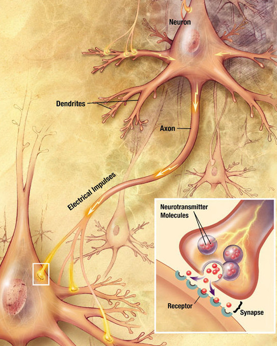
| - | Contrairement aux cellules gliales, les neurones sont capables de communiquer entre eux à travers de longues distances<ref>{{Modèle:Référence Harvard sans parenthèses | Kandel | 2000 | p=21 | id=Kandel}}</ref>. Cette communication se fait par des signaux envoyés par le biais de l'[[axone]], prolongement [[Protoplasme|protoplasmique]] du neurone qui s'étend depuis le corps cellulaire, se ramifie et se projette, parfois vers des zones proches, parfois vers des régions plus éloignées du cerveau ou du corps. Le prolongement de l'axone peut être considérable chez certains neurones. Les signaux transmis par l'axone se font sous forme d'influx électrochimiques, appelés [[Potentiel d'action|potentiels d'action]], qui durent moins d'un millième de [[seconde (temps)|seconde]] et traversent l'axone à une [[vitesse]] de 1 à 100 [[mètre]]s par seconde. Certains neurones émettent en permanence des potentiels d'action, de 10 à 100 par seconde, d'autres n'émettent des potentiels d'action qu'occasionnellement. | ||
| - | |||
| - | Le point de jonction entre l'axone d'un neurone et un autre neurone, ou une cellule non-neuronale, est la [[synapse]] où le signal est transmis<ref>{{Modèle:Référence Harvard sans parenthèses | Kandel | 2000 | loc=ch. 10 | id=Kandel}}</ref>. Un axone peut avoir jusqu'à plusieurs milliers de terminaisons synaptiques. Lorsque le potentiel d'action, après avoir parcouru l'axone, parvient à la synapse, cela provoque la libération d'un agent chimique appelé [[neurotransmetteur]]. Une fois libéré, le neurotransmetteur se lie aux [[Récepteur membranaire|récepteurs membranaires]] de la cellule cible. Certains récepteurs neuronaux sont excitateurs, c'est-à-dire qu'ils augmentent la fréquence de potentiel d'action au sein de la cellule cible ; d'autres récepteurs sont inhibiteurs et diminuent la fréquence de potentiel d'action ; d'autres ont des effets modulatoires complexes. | ||
| - | |||
| - | Les axones occupent la majeure partie de l'espace cérébral<ref>{{Modèle:Référence Harvard sans parenthèses | Kandel | 2000 | loc=ch. 2 | id=Kandel}}</ref>. Les axones sont souvent regroupés en larges groupes pour former des faisceaux de fibres nerveuses. De nombreux axones sont enveloppés d'une gaine de [[myéline]], une substance qui permet d'augmenter fortement la vitesse de propagation du potentiel d'action. La myéline est de couleur [[blanc]]he, de telle sorte que les régions du cerveau essentiellement occupées par ces fibres nerveuses apparaissent comme de la [[substance blanche]] tandis que les zones densément peuplées par les corps cellulaires des neurones apparaissent comme de la [[substance grise]]. La [[longueur]] totale des axones myélinisés dans le cerveau adulte d'un Humain dépasse en moyenne les 100 000 [[kilomètre]]s<ref>{{article|langue=en|prénom1=L.|nom1=Marner|lien auteur1=|prénom2=J. R.|nom2=Nyengaard|lien auteur2=|prénom3=Y.|nom3=Tang|lien auteur3=|prénom4=B.|nom4=Pakkenberg|lien auteur4=|traduction=|titre=Marked loss of myelinated nerve fibers in the human brain with age|sous-titre=|périodique=J. Comp. Neurol.|lien périodique=Journal of Comparative Neurology|éditeur=|lieu=|série=|volume=462|titre volume=|numéro=2|titre numéro=|jour=21|mois=juillet|année=2003|pages=144-152|issn=1096-9861|issn2=|issn3=|isbn=|résumé=|format=|url texte=|pmid=12794739|doi=10.1002/cne.10714| consulté le=|commentaire=|extrait=|id=}}</ref>. | ||
| - | |||
| - | == Développement == | ||
| - | |||
| - | [[Fichier:EmbryonicBrain.svg|thumb|Principales subdivisions du cerveau embryonnaire des Vertébrés.]] | ||
| - | Le [[développement]] du cerveau suit une succession d'étapes<ref name=purves>{{ouvrage|langue=en|prénom1=D.|nom1=Purves|lien auteur1=|directeur1=|prénom2=J. W.|nom2=Lichtman|lien auteur2=|directeur2=|prénom3=|nom3=|lien auteur3=|traducteur=|illustrateur=|préface=|titre=Principles of neural development|sous-titre=|titre original=|numéro d'édition=|collection=|série=|numéro dans collection=|lien éditeur=|éditeur=Sinauer Associates|lieu=|année=1985|mois=|jour=|année première édition=|réimpression=|tome=|volume=|titre volume=|pages totales=433|format=|isbn=9780878937448|isbn2=|isbn3=|issn=|issn2=|issn3=|oclc=|bnf=|partie=|numéro chapitre=1|titre chapitre=|passage=|présentation en ligne=http://books.google.com/books?id=t9JqAAAAMAAJ&hl=fr|lire en ligne=|consulté le=|commentaire=|extrait=|id=Purves|référence=|référence simplifié=}}</ref>. Beaucoup de neurones naissent dans des zones spécifiques contenant des [[Cellule souche|cellules souches]] et migrent ensuite à travers le tissu pour atteindre leur destination ultime<ref>{{Modèle:Référence Harvard sans parenthèses | Purves | 1985 | loc=ch. 4 |id=Purves}}</ref>. Ainsi, dans le [[Cortex cérébral|cortex]], la première étape du développement est la mise en place d'une armature par un type de [[cellule gliale|cellules gliales]], les [[cellule radiale|cellules radiales]], qui établissent des fibres verticales à travers le cortex. Les nouveaux neurones corticaux sont créés à la base du cortex et « grimpent » ensuite le long des fibres radiales jusqu'à atteindre les couches qu'ils sont destinés à occuper. | ||
| - | |||
| - | Chez les vertébrés, les premières étapes du développement sont communes à toutes les espèces<ref name=purves />. Tandis que l'embryon passe d'une forme ronde à une structure de type vermiforme, une étroite bande de l'[[ectoderme]] se décolle de la ligne médiane dorsale pour devenir la [[plaque neurale]], précurseur du [[système nerveux]]. La plaque neurale se creuse, s'[[wikt:invaginer|invagine]] de manière à former la [[gouttière neurale]] puis, les [[pli neural|plis neuraux]] qui bordent la gouttière fusionnent pour fermer la gouttière qui devient le [[tube neural]]. Ce tube se subdivise ensuite en une partie antérieure renflée, la vésicule céphalique primitive, qui se segmente en trois [[Vésicule (biologie)|vésicules]] qui deviendront le [[prosencéphale]], le [[mésencéphale]], et le [[rhombencéphale]]<ref name=purves />. Le prosencéphale se divise ensuite en deux autres vésicules, le [[télencéphale]] et le [[diencéphale]] tandis que le rhombencéphale se divise en [[métencéphale]] et [[myélencéphale]]. Chacune de ses vésicules contient des zones prolifératives dans lesquelles [[neurone]]s et [[Cellule gliale|cellules gliales]] sont formés. Ces deux types de cellules migrent ensuite, parfois sur de longues distances, vers leurs positions finales. | ||
| - | |||
| - | Une fois qu'ils sont en place, les neurones commencent à étendre leurs [[Dendrite (biologie)|dendrites]] et leur [[axone]] autour d'eux<ref>{{Modèle:Référence Harvard sans parenthèses | Purves | 1985 | loc=ch. 5 et 7 |id=Purves}}</ref>. L'axone doit généralement s'étendre sur une longue distance à partir du corps cellulaire du neurone et doit se connecter sur des cibles bien spécifiques, ce qui lui nécessite de croître d'une manière plus complexe. À l'extrémité de l'axone en développement se trouve une région parsemée de [[Récepteur (cellule)|récepteurs]] [[Chimie|chimiques]], le cône de croissance. Ces récepteurs recherchent des signaux moléculaires dans l'environnement alentour qui guident la croissance de l'axone en attirant ou en repoussant le cône de croissance et dirigent ainsi l'étirement de l'axone dans une direction donnée. Le cône de croissance navigue ainsi à travers le cerveau jusqu'à ce qu'il atteigne sa région de destination, où d'autres signaux chimiques engendrent la formation de [[synapse]]s. Des milliers de [[gène]]s interviennent pour générer ces signaux de guidage mais le réseau synaptique qui en émerge n'est déterminé qu'en partie par les [[gène]]s. Dans de nombreuses parties du cerveau, les axones connaissent d'abord une surcroissance proliférative qui est ensuite régulée par des mécanismes dépendants de l'activité neuronale<ref>{{Modèle:Référence Harvard sans parenthèses | Purves | 1985 | loc=ch. 12 |id=Purves}}</ref>. Ce processus sophistiqué de sélection et d'ajustement graduel aboutit finalement à la forme adulte du réseau neuronal. | ||
| - | |||
| - | Chez de nombreux mammifères, dont l'Homme, les neurones sont crées principalement avant la [[naissance]], et le cerveau du [[nouveau-né]] contient substantiellement plus de neurones que celui de l'[[adulte]]. A noter que notre cerveau utilise que 12,6% des neurones en moyenne. Cependant quelques zones continuent de générer de nouveaux neurones tout au long de la vie, telles que le [[bulbe olfactif]] ou le ''gyrus dentatus'' de l'[[Hippocampe (cerveau)|hippocampe]]. En dehors de ces exceptions, le nombre de neurones présents à la naissance est définitif, contrairement aux cellules gliales qui sont renouvelées tout au long de la vie, à la manière de la plupart des cellules de l'organisme. Bien que le nombre de neurones évolue peu après la naissance, les connexions axonales continuent de se développer et de s'organiser pendant encore un long moment. Chez l'Homme ce processus n'est pas terminé avant l'[[adolescence]]. | ||
| - | |||
| - | De nombreuses questions restent en suspens concernant ce qui relève de l'[[inné]] et de l'[[acquis]] à propos de l'[[esprit]], de l'[[intelligence]] et de la [[personnalité]]<ref>{{ouvrage|langue=en|prénom1=M.|nom1=Ridley|lien auteur1=|directeur1=|prénom2=|nom2=|lien auteur2=|directeur2=|prénom3=|nom3=|lien auteur3=|traducteur=|illustrateur=|préface=|titre=The agile gene|sous-titre=How nature turns on nurture|titre original=|numéro d'édition=|collection=|série=|numéro dans collection=|lien éditeur=|éditeur=Forth Estate|lieu=|année=2004|mois=|jour=|année première édition=|réimpression=|tome=|volume=|titre volume=|pages totales=352|format=|isbn=9780060006792|isbn2=|isbn3=|issn=|issn2=|issn3=|oclc=|bnf=|partie=|numéro chapitre=|titre chapitre=|passage=|présentation en ligne=http://books.google.com/books?id=9TkUHQAACAAJ&hl=fr|lire en ligne=|consulté le=|commentaire=|extrait=|id=|référence=|référence simplifié=}}</ref>. Bien que de nombreux points restent à éclaircir, la [[neuroscience]] a montré que deux facteurs sont essentiels. D'un côté, les gènes déterminent la forme générale du cerveau, et la manière dont le cerveau répond à l'expérience. D'un autre côté, l'expérience est nécessaire pour affiner la matrice de connexions synaptiques. À bien des égards, la qualité et la quantité d'expériences joue un rôle<ref>{{article|langue=en|prénom1=T. N.|nom1=Wiesel|lien auteur1=|prénom2=|nom2=|lien auteur2=|prénom3=|nom3=|lien auteur3=|traduction=|titre=Postnatal development of the visual cortex and the influence of environment|sous-titre=|périodique=Nature|lien périodique=Nature (revue)|éditeur=|lieu=|série=|volume=299|titre volume=|numéro=5884|titre numéro=|jour=14|mois=octobre|année=1982|pages=583-591|issn=0028-0836|issn2=1476-4687|issn3=|isbn=|résumé=|format=|url texte=http://nobelprize.org/nobel_prizes/medicine/laureates/1981/wiesel-lecture.pdf|pmid=6811951|doi=10.1038/299583a0| consulté le=|commentaire=|extrait=|id=}}</ref>. L’[[Enrichissement environnemental (système neural)|enrichissement environnemental]] montre que le cerveau d'un animal placé dans un environnement plus riche et stimulant a un nombre plus important de synapses que celui d'un animal dans un milieu plus pauvre<ref>{{article|langue=en|prénom1=H.|nom1=van Praag|lien auteur1=|prénom2=G.|nom2=Kempermann|lien auteur2=|prénom3=F. H.|nom3=Gage|lien auteur3=|traduction=|titre=Neural consequences of enviromental enrichment|sous-titre=|périodique=Nat. Rev. Neurosci.|lien périodique=Nature Reviews Neuroscience|éditeur=|lieu=|série=|volume=1|titre volume=|numéro=3|titre numéro=|jour=|mois=décembre|année=2000|pages=191-198|issn=1471-003X|issn2=1471-0048|issn3=|isbn=|résumé=|format=|url texte=|pmid=11257907|doi=10.1038/35044558| consulté le=|commentaire=|extrait=|id=}}</ref>. | ||
| - | |||
| - | == Fonctions == | ||
| - | |||
| - | La principale fonction du cerveau est de contrôler les actions de l'organisme à partir des [[Sens (physiologie)|informations sensorielles]] qui lui parviennent<ref>{{ouvrage|langue=en|prénom1=T. J.|nom1=Carew|lien auteur1=|directeur1=|prénom2=|nom2=|lien auteur2=|directeur2=|prénom3=|nom3=|lien auteur3=|traducteur=|illustrateur=|préface=|titre=Behavioral neurobiology|sous-titre=The cellular organization of natural behavior|titre original=|numéro d'édition=|collection=|série=|numéro dans collection=|lien éditeur=|éditeur=Sinauer Associates|lieu=|année=2000|mois=|jour=|année première édition=|réimpression=|tome=|volume=|titre volume=|pages totales=435|format=|isbn=9780878930920|isbn2=|isbn3=|issn=|issn2=|issn3=|oclc=|bnf=|partie=|numéro chapitre=1|titre chapitre=|passage=|présentation en ligne=http://books.google.com/books?id=wEMTGwAACAAJ&hl=fr|lire en ligne=|consulté le=|commentaire=|extrait=|id=|référence=|référence simplifié=}}</ref>. Les signaux sensoriels peuvent stimuler une réponse immédiate, moduler un schéma d'activité en cours, ou être emmagasinés pour un besoin futur. Ainsi, par le rôle central qu'il exerce dans la captation des [[stimulus|stimuli]] externes, le cerveau occupe le rôle central dans la création de réponses à l'environnement. Le cerveau a aussi un rôle dans la [[Hormone|régulation hormonale]]. | ||
| - | |||
| - | Le cerveau des vertébrés reçoit des signaux par les nerfs afférents de la part des différentes régions de l'organisme. Le cerveau interprète ces signaux et en tire une réponse fondée sur l'intégration des signaux électriques reçus, puis la transmet. Ce jeu de réception, d'intégration, et d'émission de signaux représente la fonction majeure du cerveau, qui explique à la fois les [[Sens (physiologie)|sensations]], le [[Mouvement (anatomie)|mouvement]], la [[mémoire (sciences humaines)|mémoire]] et, on le suppose, la [[conscience]]. | ||
| - | |||
| - | Pour mener à bien sa complexe tâche, le cerveau est organisé en sous-systèmes fonctionnels c'est-à-dire que certaines régions cérébrales traitent plus spécifiquement certains aspects de l'information. Cette division fonctionnelle n'est pas stricte et ces sous-systèmes peuvent être catégorisés de plusieurs façons : anatomiquement, chimiquement ou fonctionnellement. Une de ces catégorisations repose sur les [[neurotransmetteurs]] chimiques utilisés par les neurones pour communiquer. Une autre se base sur la manière dont chaque zone du cerveau contribue au traitement de l'information : les zones sensorielles amènent l'information au cerveau ; les signaux moteurs envoient l'information du cerveau jusqu'aux [[muscle]]s et aux [[glande]]s ; les systèmes excitateurs modulent l'activité du cerveau en fonction du moment de la journée et de divers facteurs. | ||
| - | |||
| - | Le cerveau utilise principalement le [[glucose]] comme [[Énergie|substrat énergétique]] et une [[perte de conscience]] peut survenir s'il en manque. La consommation énergétique du cerveau n'est pas particulièrement variable, mais les régions actives du cortex consomment plus d'énergie que les inactives. | ||
| - | |||
| - | === Systèmes de neurotransmissions === | ||
| - | {{Voir aussi|Neuromodulation}} | ||
| - | |||
| - | Selon le [[principe de Dale]], chaque neurone du cerveau libère constamment le même [[neurotransmetteur]] chimique, ou la même combinaison de neurotransmetteurs, pour toutes les connexions synaptiques qu'il entretient avec d'autres neurones<ref>{{Modèle:Référence Harvard sans parenthèses | Kandel | 2000 | loc=ch. 15 | id=Kandel}}</ref>. Un neurone peut donc être caractérisé en fonction des neurotransmetteurs qu'il libère bien qu'il existe quelques exceptions à ce principe. Les deux neurotransmetteurs les plus fréquents sont le [[glutamate]], qui correspond généralement à un signal excitatoire, et l'[[acide γ-aminobutyrique]] (GABA), généralement inhibitoire. Les neurones utilisant ces deux neurotransmetteurs se retrouvent dans presque toutes les régions du cerveau et forment un large pourcentage des synapses du cerveau<ref>{{ouvrage|langue=en|prénom1=P. L.|nom1=McGeer|lien auteur1=|directeur1=|prénom2=E. G.|nom2=McGeer|lien auteur2=|directeur2=|prénom3=|nom3=|lien auteur3=|traducteur=|illustrateur=|préface=|titre=Basic neurochemistry|sous-titre=Molecular, cellular and medical aspects |titre original=|numéro d'édition=4|collection=|série=|numéro dans collection=|lien éditeur=|éditeur=Raven Press|lieu=New York|année=1989|mois=|jour=|année première édition=|réimpression=|tome=|volume=|titre volume=|pages totales=984|format=|isbn=9780881673432|isbn2=|isbn3=|issn=|issn2=|issn3=|oclc=|bnf=|partie=|numéro chapitre=15|titre chapitre=Amino acid neurotransmitters|passage=311-332|présentation en ligne=http://books.google.com/books?id=b-FqAAAAMAAJ&q=isbn:9780881673432&dq=isbn:9780881673432&hl=fr&ei=8E5JTYuLBMO24QaHyeXdCw&sa=X&oi=book_result&ct=result&resnum=1&ved=0CCkQ6AEwAA|lire en ligne=|consulté le=|commentaire=|extrait=|id=|référence=|référence simplifié=}}</ref>. | ||
| - | |||
| - | Les autres neurotransmetteurs, comme la [[sérotonine]] ou la [[noradrénaline]], proviennent de neurones localisés dans des zones particulières du cerveau. D'autres neurotransmetteurs, comme l'[[acétylcholine]] ou la [[dopamine]], proviennent de plusieurs endroits du cerveau, mais ne sont pas distribués de façon aussi ubiquitaire que le glutamate et le GABA. La grande majorité des [[drogue]]s [[psychotrope]]s agissent en altérant les systèmes de neurotransmetteurs qui ne sont pas directement impliqués dans les transmissions glutamatergiques ou GABAergiques<ref>{{ouvrage|langue=en|prénom1=J. R.|nom1=Cooper|lien auteur1=|directeur1=|prénom2=F. E.|nom2=Bloom|lien auteur2=|directeur2=|prénom3=R. H.|nom3=Roth|lien auteur3=|traducteur=|illustrateur=|préface=|titre=The biochemical basis of neuropharmacology|sous-titre=|titre original=|numéro d'édition=8|collection=|série=|numéro dans collection=|lien éditeur=Oxford University Press|éditeur=Oxford University Press|lieu=|année=2003|mois=|jour=|année première édition=|réimpression=|tome=|volume=|titre volume=|pages totales=405|format=|isbn=9780195140088|isbn2=|isbn3=|issn=|issn2=|issn3=|oclc=|bnf=|partie=|numéro chapitre=|titre chapitre=|passage=|présentation en ligne=http://books.google.com/books?id=e5I5gOwxVMkC&hl=fr|lire en ligne=|consulté le=|commentaire=|extrait=|id=|référence=|référence simplifié=}}</ref>. | ||
| - | |||
| - | === Systèmes sensoriels === | ||
| - | {{Voir aussi|Système sensoriel}} | ||
| - | |||
| - | Une fonction importante du cerveau est de traiter l'information reçue par les [[Récepteur sensoriel|récepteurs sensoriels]]<ref>{{Modèle:Référence Harvard sans parenthèses | Kandel | 2000 | loc=ch. 21 | id=Kandel}}</ref>. Contrairement aux idées reçues, les [[Sens (physiologie)|sens]] que peut capter le cerveau ne sont pas limitées à cinq. Outre la [[vue]], l'[[ouïe]], le [[toucher]], l'[[odorat]], et le [[goût]], le cerveau peut recevoir d'autres informations sensorielles comme la [[température]], l'[[équilibre]], la [[position]] des membres, ou la [[composition chimique]] du [[sang]]. Toutes ces variables sont détectées par des récepteurs spécialisés qui transmettent les signaux vers le cerveau. Certaines espèces peuvent détecter des sens supplémentaires, comme la vision [[infrarouge]] des [[serpent]]s, ou utiliser les sens « standards » de manière non conventionnelle, comme l'[[écholocation]] du système auditif des [[chauves-souris]]. | ||
| - | |||
| - | Chaque système sensoriel possède ses propres cellules sensorielles réceptrices. Ces cellules sont des neurones mais, contrairement à la majorité des neurones, ceux-ci ne sont pas contrôlés par les signaux synaptiques d'autres neurones. Au lieu de cela, ces cellules sensorielles possèdent des [[Récepteur membranaire|récepteurs membranaires]] qui sont stimulées par un facteur physique spécifique comme la [[lumière]], la [[température]], ou la [[pression]]. Les signaux de ces cellules sensorielles réceptrices parviennent jusqu'à la [[moelle épinière]] ou le cerveau par les [[nerf]]s afférents. | ||
| - | |||
| - | Pour la plupart des sens, il y a un noyau sensitif principal dans le [[tronc cérébral]], ou un ensemble de noyaux, qui reçoit et réunit les signaux des cellules sensorielles réceptrices. Dans de nombreux cas, des zones secondaires sous-corticales se chargent d'extraire et de trier l'information. Chaque système sensoriel a également une région du [[thalamus]] qui lui est dédié et qui relaie l'information au [[cortex]]. | ||
| - | |||
| - | Pour chaque système sensoriel, une zone corticale primaire reçoit directement les signaux en provenance du relai thalamique. Habituellement, un groupe spécifique de zones corticales supérieures analyse également le signal sensoriel. Enfin, des zones multimodales du cortex combinent les signaux en provenance de différents systèmes sensoriels. À ce niveau, les signaux qui atteignent ces régions du cerveau sont considérés comme des signaux intégrés plutôt que comme des signaux strictement sensoriels<ref>{{Modèle:Référence Harvard sans parenthèses | Kandel | 2000 | loc=ch. 21 et 30 | id=Kandel}}</ref>. | ||
| - | |||
| - | Toutes ces étapes ont leurs exceptions. Ainsi, pour le toucher, les signaux sensoriels sont principalement reçus au niveau de la moelle épinière, au niveau de neurones qui projettent ensuite l'information au tronc cérébral<ref>{{Modèle:Référence Harvard sans parenthèses | Kandel | 2000 | loc=ch. 23 | id=Kandel}}</ref>. Pour l'odorat, il n'y a pas de relai dans le thalamus, le signal est transmis directement de la zone primaire, le [[bulbe olfactif]], vers le cortex<ref>{{Modèle:Référence Harvard sans parenthèses | Kandel | 2000 | loc=ch. 32 | id=Kandel}}</ref>. | ||
| - | |||
| - | === Systèmes moteurs === | ||
| - | |||
| - | Les systèmes moteurs sont les zones du cerveau responsables directement ou indirectement des mouvements du corps, en agissant sur les [[muscle]]s. À l'exception des muscles contrôlant les [[yeux]], tous les [[Muscle strié|muscles striés]] de l'organisme sont directement innervés par des [[Motoneurone|neurones moteurs]] de la [[moelle épinière]]. Ils sont donc le dernier maillon de la chaîne du système psycho-moteur<ref>{{Modèle:Référence Harvard sans parenthèses | Kandel | 2000 | loc=ch. 34 | id=Kandel}}</ref>. Les neurones moteurs spinaux sont contrôlés à la fois par des circuits neuronaux propres à la moelle épinière, et par des influx efférents du cerveau. Les circuits spinaux intrinsèques hébergent plusieurs [[réflexe (réaction motrice)|réactions réflexes]], ainsi que certains schémas de mouvements comme les mouvements rythmiques tels que la [[marche]] ou la [[nage]]<ref>{{Modèle:Référence Harvard sans parenthèses | Kandel | 2000 | loc=ch. 36-37 | id=Kandel}}</ref>. Les connexions efférentes du cerveau permettent quant à elles, des contrôles plus sophistiqués. | ||
| - | |||
| - | Un certain nombre de zones du cerveau sont connectées directement à la moelle épinière<ref>{{Modèle:Référence Harvard sans parenthèses | Kandel | 2000 | loc=ch. 33 | id=Kandel}}</ref>. Au niveau le plus inférieur se trouve les zones moteurs situées dans le [[bulbe rachidien]] et le [[Pont (système nerveux)|pont]]. Au-dessus se situent les zones du mésencéphale, comme le [[noyau rouge]], qui sont responsables de la coordination des mouvements. À un niveau supérieur se trouve le [[cortex moteur primaire]], une bande de tissu cérébral localisé à la lisière postérieure du lobe frontal. Le cortex moteur primaire transmet ses commandes motrices aux zones moteurs sous-corticales, mais également directement à la moelle épinière par le biais du [[faisceau pyramidal]]. Les influx nerveux de ce faisceau cortico-spinal transmettent les mouvements fins volontaires. D'autres zones moteurs du cerveau ne sont pas directement reliées à la moelle épinière, mais agissent sur les zones moteurs primaires corticales ou sous-corticales. Quelques une de ces zones secondaires les plus importantes sont le [[cortex prémoteur]], impliqués dans la coordination des mouvements de différentes parties du corps, les [[ganglions de la base]], dont la fonction principale semble être la sélection de l'action, et le [[cervelet]], qui module et optimise les informations pour rendre les mouvements plus précis. | ||
| - | |||
| - | Le cerveau et la moelle épinière contiennent également un réseau neuronal qui contrôle le [[système nerveux autonome]], la partie du système nerveux responsable des fonctions automatiques. Non soumis au contrôle volontaire, le système nerveux autonome contrôle notamment la [[Hormone|régulation hormonale]] et l'activité des [[muscle lisse|muscles lisses]] et du [[muscle cardiaque]]. Le système nerveux autonome agit à différents niveaux comme le [[rythme cardiaque]], la [[digestion]], la [[respiration]], la [[salivation]], la [[miction]], la [[sueur]] ou l'[[excitation sexuelle]]. | ||
| - | |||
| - | === Systèmes d'éveil === | ||
| - | |||
| - | Un des aspects les plus visibles du comportement animal est le cycle journalier veille-sommeil-rêve. L'éveil et l'attention sont aussi modulés à une échelle de temps plus fine, par un réseau de zones cérébrales<ref name="neural45">{{Modèle:Référence Harvard sans parenthèses | Kandel | 2000 | loc=ch. 45 | id=Kandel}}</ref>. | ||
| - | |||
| - | Un composant clé du système d'éveil est le [[noyau suprachiasmatique]], petite région de l'[[hypothalamus]] localisée directement au-dessus du point de croisement des [[nerf optique|nerfs optiques]]<ref>{{article|langue=en|prénom1=M. C.|nom1=Antle|lien auteur1=|prénom2=R.|nom2=Silver|lien auteur2=|prénom3=|nom3=|lien auteur3=|traduction=|titre=Orchestrating time: arrangements of the brain circadian clock|sous-titre=|périodique=Trends Neurosci.|lien périodique=Trends in Neurosciences|éditeur=|lieu=|série=|volume=28|titre volume=|numéro=3|titre numéro=|jour=|mois=mars|année=2005|pages=145-151|issn=0166-2236|issn2=|issn3=|isbn=|résumé=|format=|url texte=http://www.columbia.edu/cu/psychology/silver/publications2/149%20antle%20et%20al.pdf|pmid=15749168 |doi=10.1016/j.tins.2005.01.003| consulté le=|commentaire=|extrait=|id=}}</ref>. Le noyau suprachiasmatique renferme l'[[Horloge circadienne|horloge biologique]] centrale de l'organisme. Les neurones de ce noyau montrent un niveau d'activité qui augmente ou diminue sur une période d'environ 24 heures, le [[rythme circadien]] : cette activité fluctuante est dirigée par des changements rythmiques exprimés par un groupe de gènes horlogers. Le noyau suprachiasmatique reçoit généralement des signaux en provenance des nerfs optiques qui permettent de calibrer l'horloge biologique à partir des cycles jour-nuit. | ||
| - | |||
| - | Le noyau suprachiasmatique se projette dans un ensembles de zones cérébrales, situées au niveau de l'[[hypothalamus]], du [[tronc cérébral]], et du [[mésencéphale]], qui sont impliqués dans la mise en œuvre des cycles jour-nuit. Un composant important du système est la [[formation réticulée]], un groupe d'amas neuronaux s'étendant dans le tronc cérébral<ref name="neural45"/>. Les neurones réticulés envoient des signaux vers le [[thalamus]], qui répond en envoyant des signaux à différentes régions du [[cortex]] qui régule le niveau d'activité. | ||
| - | |||
| - | Le [[sommeil]] implique de profondes modifications dans l'activité cérébrale<ref>{{Modèle:Référence Harvard sans parenthèses | Kandel | 2000 | loc=ch. 47 | id=Kandel}}</ref>. Le cerveau ne s'éteint pas pendant le sommeil, l'activité cérébrale se poursuit mais est modifiée. En fait, il existe deux types de sommeil : le [[sommeil paradoxal]] (avec [[rêve]]s) et le sommeil non paradoxal (généralement sans rêves). Ces deux sommeils se répètent selon un schéma légèrement différent à chaque sommeil. Trois grands types de schéma d'activité cérébrale peuvent être distingués : sommeil paradoxal, sommeil léger, et sommeil profond. Pendant le sommeil profond, l'activité du cortex prend la forme de larges ondes synchronisées tandis que ces ondes sont désynchronisées pendant l'état de rêve. Les niveaux de [[noradrénaline]] et de [[sérotonine]] tombent au cours du sommeil profond, et approchent du niveau zéro pendant le sommeil paradoxal, tandis que les niveaux d'[[acétylcholine]] présentent un schéma inverse. | ||
| - | |||
| - | == Le cerveau et l'esprit == | ||
| - | {{Voir aussi|Philosophie de l'esprit}} | ||
| - | |||
| - | La compréhension de la relation entre le cerveau et l'[[esprit]] est un problème aussi bien [[scientifique]] que [[philosophique]]<ref>{{ouvrage|langue=en|prénom1=P. S|nom1=Churchland|lien auteur1=|directeur1=|prénom2=|nom2=|lien auteur2=|directeur2=|prénom3=|nom3=|lien auteur3=|traducteur=|illustrateur=|préface=|titre=Neurophilosophy|sous-titre=Toward a unified science of the mind-brain|titre original=|numéro d'édition=5|collection=|série=|numéro dans collection=|lien éditeur=|éditeur=MIT Press|lieu=|année=1990|mois=|jour=|année première édition=|réimpression=|tome=|volume=|titre volume=|pages totales=546|format=|isbn=9780262530859|isbn2=|isbn3=|issn=|issn2=|issn3=|oclc=|bnf=|partie=|numéro chapitre=|titre chapitre=|passage=|présentation en ligne=http://books.google.com/books?id=hAeFMFW3rDUC&hl=fr|lire en ligne=|consulté le=|commentaire=|extrait=|id=Churchland|référence=|référence simplifié=}}</ref>. La relation forte entre la matière cérébrale physique et l'esprit est aisément mise en évidence par l'impact que les altérations physiques du cerveau ont sur l'esprit, comme le [[traumatisme crânien]] ou l'usage de [[psychotrope]]<ref>{{ouvrage|langue=en|prénom1=C.|nom1=Boake|lien auteur1=|directeur1=|prénom2=L.|nom2=Dillar|lien auteur2=|directeur2=|prénom3=|nom3=|lien auteur3=|traducteur=|illustrateur=|préface=|titre=Rehabilitation for Traumatic Brain Injury|sous-titre=|titre original=|numéro d'édition=|collection=|série=|numéro dans collection=|lien éditeur=Oxford University Press|éditeur=Oxford University Press|lieu=|année=2005|mois=juin|jour=|année première édition=|réimpression=|tome=|volume=|titre volume=|pages totales=385|format=|isbn=9780195173550|isbn2=|isbn3=|issn=|issn2=|issn3=|oclc=|bnf=|partie=|numéro chapitre=1|titre chapitre=History of Rehabilitation for Traumatic Brain Injury|passage=|présentation en ligne=|lire en ligne=|consulté le=|commentaire=|extrait=|id=|référence=|référence simplifié=}}</ref>. | ||
| - | |||
| - | Le [[problème corps-esprit]] est l'un des débat centraux de l'[[histoire de la philosophie]] et consiste à considérer la manière dont le cerveau et l'esprit sont reliés<ref>{{Modèle:Référence Harvard sans parenthèses | Churchland| 1990| loc=ch. 7|id=Churchland}}</ref>. Trois grands courants de pensée existent concernant cette question : [[Dualisme (philosophie de l'esprit)|dualisme]], [[matérialisme]], et [[idéalisme]]. Le dualisme postule que l'esprit existe indépendamment du cerveau ; le matérialisme postule, quant à lui, que le phénomène mental est identique au phénomène neuronal ; et l'idéalisme postule que seul le phénomène mental existe<ref>{{ouvrage|langue=en|prénom1=W. D.|nom1=Hart|lien auteur1=|directeur1=|prénom2=S.|nom2=Guttenplan|lien auteur2=|directeur2=oui|prénom3=|nom3=|lien auteur3=|traducteur=|illustrateur=|préface=|titre=A companion to the philosophy of mind|sous-titre=|titre original=|numéro d'édition=|collection=|série=|numéro dans collection=|lien éditeur=John Wiley & Sons|éditeur=Wiley-Blackwell|lieu=|année=1996|mois=|jour=|année première édition=|réimpression=|tome=|volume=|titre volume=|pages totales=656|format=|isbn=9780631199960|isbn2=|isbn3=|issn=|issn2=|issn3=|oclc=|bnf=|partie=|numéro chapitre=|titre chapitre=Dualism|passage=265-267|présentation en ligne=|lire en ligne=http://books.google.fr/books?id=GFtVBGAn5-YC&lpg=PP1&dq=9780631199960&pg=PA265#v=onepage&q&f=false|consulté le=|commentaire=|extrait=|id=|référence=|référence simplifié=}}</ref>{{,}}<ref>{{ouvrage|langue=en|prénom1=A. R.|nom1=Lacey|lien auteur1=|directeur1=|prénom2=|nom2=|lien auteur2=|directeur2=|prénom3=|nom3=|lien auteur3=|traducteur=|illustrateur=|préface=|titre=A dictionary of philosophy|sous-titre=|titre original=|numéro d'édition=3|collection=|série=|numéro dans collection=|lien éditeur=Routledge|éditeur=Routledge|lieu=|année=1996|mois=|jour=|année première édition=1976|réimpression=|tome=|volume=|titre volume=|pages totales=386|format=|isbn=9780415133326|isbn2=|isbn3=|issn=|issn2=|issn3=|oclc=|bnf=|partie=|numéro chapitre=|titre chapitre=|passage=|présentation en ligne=|lire en ligne=http://books.google.fr/books?id=kJaCVLZrBcoC&lpg=PP1&ots=Bfyl2mly-x&dq=%22A%20Dictionary%20of%20Philosophy%22%20lacey&pg=PP1#v=onepage&q&f=false|consulté le=|commentaire=|extrait=|id=|référence=|référence simplifié=}}</ref>. | ||
| - | |||
| - | Outre ces questions philosophiques, la relation entre l'esprit et le cerveau soulève un grand nombre de questions scientifiques, comme la relation entre l'activité mentale et l'activité cérébrale, le mécanisme d'action des drogues sur la [[cognition]], ou encore la corrélation entre neurones et [[conscience]]. | ||
| - | |||
| - | Historiquement, un grand nombre de philosophes considéraient inconcevable que la cognition puisse être mise en place par une substance physique comme le tissu cérébral<ref>{{Modèle:Référence Harvard sans parenthèses | Churchland| 1990| loc=ch. 6|id=Churchland}}</ref>. Des philosophes comme [[Patricia Churchland]] ont postulé que l'interaction entre la drogue et l'esprit est un indicateur de la relation intime entre le cerveau et l'esprit mais que les deux entités sont distinctes<ref>{{Modèle:Référence Harvard sans parenthèses | Churchland| 1990| loc=ch. 8|id=Churchland}}</ref>. [[René Descartes]] trouvait qu'il était possible d'expliquer les réflexes et d'autres comportements simples en termes mécaniques, mais il ne croyait pas que la pensée complexe, notamment le langage, puisse être expliqué par le cerveau physique uniquement<ref>{{ouvrage|langue=en|prénom1=R.|nom1=Descartes|lien auteur1=René Descartes|directeur1=|prénom2=|nom2=|lien auteur2=|directeur2=|prénom3=|nom3=|lien auteur3=|traducteur=R. Stoothoff, D. Murdoch, J. Cottingham|illustrateur=|préface=|titre=The philosophical writings of Descartes|sous-titre=|titre original=|numéro d'édition=|collection=|série=|numéro dans collection=|lien éditeur=Cambridge University Press|éditeur=Cambridge University Press|lieu=|année=1991|mois=|jour=|année première édition=|réimpression=|tome=|volume=|titre volume=|pages totales=448|format=|isbn=9780521288088|isbn2=|isbn3=|issn=|issn2=|issn3=|oclc=|bnf=|partie=|numéro chapitre=|titre chapitre=|passage=|présentation en ligne=|lire en ligne=|consulté le=|commentaire=|extrait=|id=|référence=|référence simplifié=}}</ref>. | ||
| - | |||
| - | == Recherche scientifique == | ||
| - | {{Voir aussi|Neurosciences}} | ||
| - | [[Fichier:PET-image.jpg|thumb|Vue axiale du cerveau par [[tomographie par émission de positons]].]] | ||
| - | |||
| - | Le domaine des [[neurosciences]] englobe toutes les approches cherchant à comprendre le fonctionnement du cerveau et du reste du [[système nerveux]]<ref>{{Modèle:Référence Harvard sans parenthèses | Kandel | 2000 | loc=ch. 1| id=Kandel}}</ref>. La psychologie cherche à comprendre l'[[esprit]] et le [[comportement]]. La [[neurologie]] est la [[Médecine|discipline médicale]] qui diagnostique et traite les [[pathologie]]s liées au système nerveux. Le cerveau est également l'organe le plus étudié en [[psychiatrie]], une branche de la médecine qui étudie et traite les [[Maladie mentale|troubles mentaux]]<ref>{{ouvrage|langue=en|prénom1=H. A.|nom1=Storrow|lien auteur1=|directeur1=|prénom2=|nom2=|lien auteur2=|directeur2=|prénom3=|nom3=|lien auteur3=|traducteur=|illustrateur=|préface=|titre=Outline of clinical psychiatry|sous-titre=|titre original=|numéro d'édition=|collection=|série=|numéro dans collection=|lien éditeur=|éditeur=Appleton-Century-Crofts|lieu=|année=1969|mois=|jour=|année première édition=|réimpression=|tome=|volume=|titre volume=|pages totales=|format=|isbn=9780390850751|isbn2=|isbn3=|issn=|issn2=|issn3=|oclc=|bnf=|partie=|numéro chapitre=|titre chapitre=|passage=|présentation en ligne=|lire en ligne=|consulté le=|commentaire=|extrait=|id=|référence=|référence simplifié=}}</ref>. Les [[sciences cognitives]] tentent de lier la neuroscience et la psychologie avec d'autres domaines comme l'[[informatique]] et la [[philosophie]]. | ||
| - | |||
| - | La plus ancienne méthode d'étude du cerveau est l'[[anatomie]]. Au milieu du [[XXe siècle|XX{{e}} siècle]], les progrès des neurosciences proviennent de l'amélioration des techniques de [[microscopie]] et de [[Coloration (histologie)|coloration]]<ref>{{ouvrage|langue=en|prénom1=M. F.|nom1=Bear|lien auteur1=|directeur1=|prénom2=B. W.|nom2=Connors|lien auteur2=|directeur2=|prénom3=M. A.|nom3=Paradiso|lien auteur3=|traducteur=|illustrateur=|préface=|titre=Neuroscience|sous-titre=Exploring the brain|titre original=|numéro d'édition=3|collection=|série=|numéro dans collection=|lien éditeur=|éditeur=Lippincott Williams & Wilkins|lieu=|année=|mois=|jour=|année première édition=|réimpression=|tome=|volume=|titre volume=|pages totales=857|format=|isbn=9780781760034|isbn2=|isbn3=|issn=|issn2=|issn3=|oclc=|bnf=|partie=|numéro chapitre=2|titre chapitre=Neurons and glia|passage=|présentation en ligne=|lire en ligne=http://books.google.fr/books?id=DbahEn-y6AoC&lpg=PA23&dq=%22Neuroscience%3A%20Exploring%20the%20Brain%22&pg=PA23#v=onepage&q&f=false|consulté le=|commentaire=|extrait=|id=|référence=|référence simplifié=}}</ref>. Les neuroanatomistes étudient la structure du cerveau aussi bien à grande échelle qu'à l'échelle microscopique. Parmi d'autres outils, ils emploient une large gamme de colorants qui permettent de révéler la structure neurale, les réactions chimiques, et la connectivité. Le développement plus récent de techniques d'[[immunocoloration]] a permis de colorer les neurones qui exprime spécifiquement un groupe de [[gène]]s. Également, la neuroanatomie fonctionnelle utilise les techniques d'[[imagerie médicale]] pour corréler les variations dans la structure du cerveau avec les changements de [[cognition]] ou de [[comportement]]. | ||
| - | |||
| - | Les neurophysiologistes étudient les propriétés chimiques, pharmacologiques et électriques du cerveau. Leur principaux outils sont les drogues et les dispositifs d'enregistrement. Des milliers de drogues expérimentalement développées affectent le système nerveux, plusieurs le font de manière très spécifique. L'enregistrement de l'activité cérébrale peut se faire par l'utilisation d'[[électrode]]s, soit collées au [[crâne]] comme dans le cas d'[[électro-encéphalographie]], soit implantées à l'intérieur du cerveau pour des enregistrements extracellulaires, qui peuvent détecter les [[Potentiel d'action|potentiels d'action]] générés par des neurones individuels. Comme le cerveau ne contient pas de [[nocicepteur]]s, il est possible d'utiliser ces techniques sur un animal éveillé sans causer de [[douleur]]. Il est aussi possible d'étudier l'activité cérébrale par un [[examen non invasif]] en utilisant des techniques d'imagerie fonctionnelle comme l'[[Imagerie par résonance magnétique|IRM]]. Ainsi la [[tomographie à émission de positons]] met en évidence qu'en l'absence de toute focalisation particulière de l'attention, l'activité du cerveau (activité intrinsèque du réseau du mode par défaut, nommée « énergie sombre du cerveau » par analogie à l'[[énergie sombre]] du cosmos<ref>{{Article | ||
| - | |langue=en|nom1=Marcus Raichle|titre=The Brain's Dark Energy|périodique=Science|année=2009|volume=314|pages=1249-1250}}</ref> et qui consiste en des vagues d'ondes électriques lentes) correspond à une dépense de 60 à 80% de toute l'énergie consommée par le cerveau, soit une énergie 20 fois supérieure à celle consommée par le cerveau lorsqu'il réagit consciemment, la réalisation d'une tâche particulière (activite consciente moins fréquente que l'activité inconsciente) exige une énergie qui n'excède pas 5% de celle consommée par l'activité de fond<ref>{{Article|nom1={{Lien|Marcus Raichle}}|titre=Un cerveau jamais au repos|périodique=Pour la Science|année=2010|numéro=393|pages=42-47}}</ref>. | ||
| - | |||
| - | Une autre approche est d'examiner les conséquences de l'endommagement de zones spécifiques du cerveau. Bien que protégé par le crâne et les méninges, et isolé du flux sanguin par la [[Barrière hémato-encéphalique]], le cerveau est tout de même vulnérable à de nombreuses maladies et à différents types de dégâts. Chez les humains, les effets des dégâts cérébraux sont une source importante d'informations sur la fonction cérébrale<ref>{{ouvrage|langue=en|prénom1=B.|nom1=Kolb|lien auteur1=|directeur1=|prénom2=I. Q.|nom2=Whishaw|lien auteur2=|directeur2=|prénom3=|nom3=|lien auteur3=|traducteur=|illustrateur=|préface=|titre=Fundamentals of human neuropsychology|sous-titre=|titre original=|numéro d'édition=6|collection=|série=|numéro dans collection=|lien éditeur=|éditeur=Worth Publisherslieu=|année=2008|mois=|jour=|année première édition=|réimpression=|tome=|volume=|titre volume=|pages totales=763|format=|isbn=9780716795865|isbn2=|isbn3=|issn=|issn2=|issn3=|oclc=|bnf=|partie=|numéro chapitre=1|titre chapitre=|passage=|présentation en ligne=http://books.google.fr/books?id=z0DThNQqdL4C|lire en ligne=|consulté le=|commentaire=|extrait=|id=|référence=|référence simplifié=}}</ref>. Comme il n'y a pas la capacité de contrôler expérimentalement la nature de ces dégâts, cette information est néanmoins souvent difficile à interpréter. Chez les animaux, les [[rat]]s étant les plus fréquents sujets d'étude, il est possible d'utiliser des électrodes ou d'injecter localement des produits chimiques pour produire des types de dégâts bien précis et observer ensuite leurs conséquences sur le comportement. | ||
| - | |||
| - | Les [[neurosciences computationnelles]] regroupent deux approches : l'utilisation d'ordinateurs pour comprendre le cerveau et l'étude de la façon dont le cerveau réalise la [[computation]]<ref>{{ouvrage|langue=en|prénom1=P.|nom1=Dayan|lien auteur1=|directeur1=|prénom2=L. F.|nom2=Abbott|lien auteur2=|directeur2=|prénom3=|nom3=|lien auteur3=|traducteur=|illustrateur=|préface=|titre=Theoretical neuroscience|sous-titre=Computational and mathematical modeling of neural systems|titre original=|numéro d'édition=|collection=|série=|numéro dans collection=|lien éditeur=MIT Press|éditeur=MIT Press|lieu=|année=2005|mois=|jour=|année première édition=|réimpression=|tome=|volume=|titre volume=|pages totales=460|format=|isbn=9780262541855|isbn2=|isbn3=|issn=|issn2=|issn3=|oclc=|bnf=|partie=|numéro chapitre=|titre chapitre=|passage=|présentation en ligne=http://books.google.com/books?id=hrZYAAAACAAJ&hl=fr|lire en ligne=|consulté le=|commentaire=|extrait=|id=|référence=|référence simplifié=}}</ref>. D'un côté, il est possible de [[Codage (programmation)|coder]] un [[programme informatique]] pour permettre de [[Simulation informatique|simuler]] le fonctionnement d'un groupe de neurones en utilisant des systèmes d'équations décrivant l'activité électrochimique ; ces simulations sont appelées « réseaux de neurones biologiquement réalistes ». D'un autre côté, il est possible d'étudier les [[algorithmes]] de computation neurale par la [[simulation]] ou l'[[analyse mathématique]] d'« unités » simplifiées ayant plusieurs des caractéristiques des neurones mais en faisant abstraction de la plupart de leur complexité biologique. Les fonctions computationnelles du cerveau sont étudiés à la fois par les neuroscientifiques et les informaticiens. | ||
| - | |||
| - | Les dernières années ont vu les premières applications des techniques d'ingénierie génétique pour l'étude du cerveau<ref>{{article|langue=en|prénom1=S.|nom1=Tonegawa|lien auteur1=|prénom2=K.|nom2=Nakazawa|lien auteur2=|prénom3=M. A.|nom3=Wilson|lien auteur3=|traduction=|titre=Genetic neuroscience of mammalian learning and memory|sous-titre=|périodique=Phil. Trans. R. Soc. B|lien périodique=Philosophical Transactions of the Royal Society B: Biological Sciences|éditeur=|lieu=|série=|volume=358|titre volume=|numéro=1432|titre numéro=|jour=29|mois=avril|année=2003|pages=787-795|issn=1471-2970|issn2=|issn3=|isbn=|résumé=|format=|url texte=|pmid=12740125|doi=10.1098/rstb.2002.1243| consulté le=|commentaire=|extrait=|id=}}</ref>. Les sujets d'études les plus fréquents sont les souris, car c'est sur cette espèce que les outils techniques à disposition sont le plus au point. Il est désormais possible d'inactiver ou de muter une grande variété de gènes, et ensuite examiner les effets sur la fonction cérébrale. Des approches plus sophistiquées sont également utilisées, comme la [[recombinaison Cre-Lox]] qui permet d'activer ou d'inactiver les gènes dans des zones spécifiques du cerveau à des moments spécifiques. | ||
| - | |||
| - | Des équipes américaines, bénéficiant d'un financement fédéral de 28 millions d'euros, utilisent des [[scanner]]s dernier cri pour créer une banque de « connectomes ». Ces cartes des circuits cérébraux promettent de révéler comment les organes réagissent au vieillissement, à l'apprentissage et à d'autres événements. Les données du [[Human Connectome Project]] laisseraient présager des avancées sur les traitements de l'[[autisme]] et de la [[schizophrénie]]. | ||
| - | |||
| - | == Histoire == | ||
| - | {{Voir aussi|Histoire de la connaissance du cerveau}} | ||
| - | |||
| - | Historiquement, les avis se sont souvent opposés pour savoir qui du cerveau ou du [[cœur]] était le siège de l'[[âme]]. Dans un sens, il était impossible de nier que la conscience semble comme localisée dans la [[tête (anatomie)|tête]], qu'un coup à la tête provoque bien plus facilement l'inconscience qu'un coup sur le [[torse]], et que secouer la tête donne des [[vertige]]s. Dans un autre sens, le cerveau soumis à un examen superficiel semble inerte, tandis que le cœur bat constamment. L'arrêt des [[Battement cardiaque|battements cardiaques]] provoque la mort, tandis que les émotions induisent des changements du [[rythme cardiaque]] et que le chagrin produit souvent une sensation de douleur au niveau du cœur (« cœur brisé »). Pour [[Aristote]], le siège de l'âme était le cœur et le cerveau, organe de refroidissement, servait simplement à assurer la [[circulation du sang]] (théorie philosophique et médicale du cardiocentrisme). [[Démocrite]] divise l'âme en trois parties : l'intellect dans la tête, l'émotion dans le cœur, le désir autour du [[foie]]<ref>{{ouvrage|langue=en|prénom1=S.|nom1=Finger|lien auteur1=|directeur1=|prénom2=|nom2=|lien auteur2=|directeur2=|prénom3=|nom3=|lien auteur3=|traducteur=|illustrateur=|préface=|titre=Origins of neuroscience|sous-titre=A history of explorations into brain function|titre original=|numéro d'édition=|collection=|série=|numéro dans collection=|lien éditeur=Oxford University Press|éditeur=Oxford University Press|lieu=|année=2001|mois=|jour=|année première édition=|réimpression=|tome=|volume=|titre volume=|pages totales=480|format=|isbn=9780195146943|isbn2=|isbn3=|issn=|issn2=|issn3=|oclc=|bnf=|partie=|numéro chapitre=|titre chapitre=|passage=14|présentation en ligne=http://books.google.com/books?id=_GMeW9E1IB4C&hl=fr|lire en ligne=|consulté le=|commentaire=|extrait=|id=|référence=|référence simplifié=}}</ref>. [[Hippocrate]] était sûr que l'âme se trouvait dans le cerveau (théorie philosophique et médicale du cérébrocentrisme). Pour [[Hérophile]], le cerveau était le centre de l’intelligence<ref>[http://psychobiologierouen.free.fr/document/S1UE2.pdf De la psychobiologie aux neurosciences cognitives]{{pdf}}</ref>. | ||
| - | |||
| - | [[Fichier:PurkinjeCell.jpg|thumb|Dessin de Santiago Ramón y Cajal de neurones de pigeon observés grâce à la coloration de Golgi.]] | ||
| - | |||
| - | [[Galien]] a aussi défendu l'importance du cerveau, et élaboré des théories assez avancées sur sa manière de fonctionner. Même après la reconnaissance de la primauté du cerveau par la [[société savante]], l'idée du cœur comme siège de l'intelligence a continué de survivre dans les [[idiome]]s populaires<ref>{{ouvrage|langue=en|prénom1=R.|nom1=Hendrickson|lien auteur1=|directeur1=|prénom2=|nom2=|lien auteur2=|directeur2=|prénom3=|nom3=|lien auteur3=|traducteur=|illustrateur=|préface=|titre=The facts on file encyclopedia of word and phrase origins|sous-titre=|titre original=|numéro d'édition=4|collection=|série=|numéro dans collection=|lien éditeur=|éditeur=Facts on File|lieu=New York|année=2008|mois=|jour=|année première édition=|réimpression=|tome=|volume=|titre volume=|pages totales=948|format=|isbn=9780816069668|isbn2=|isbn3=|issn=|issn2=|issn3=|oclc=|bnf=|partie=|numéro chapitre=|titre chapitre=|passage=|présentation en ligne=http://books.google.com/books?id=JZfyGgAACAAJ|lire en ligne=|consulté le=|commentaire=|extrait=|id=|référence=|référence simplifié=}}</ref>. Galien a entrepris un long travail pour décrire les relations anatomiques entre le cerveau, les [[nerf]]s, et les [[muscle]]s, démontrant que tous les muscles du corps sont connectés au cerveau par un réseau de nerfs. Il postulait que les nerfs activent les muscles mécaniquement, en transportant une substance mystérieuse qu'il appelle le pneuma psychique (ou « esprit animal »). Ses idées connaissent une relative renommée pendant le [[Moyen Âge|Moyen-Âge]], mais ne progressent pas jusqu'à la [[Renaissance (période historique)|Renaissance]]. À la Renaissance, des études anatomiques détaillées résument et combinent les idées de Galien avec celles de [[Descartes]] et ses successeurs. Comme Galien, Descartes pensait le [[système nerveux]] en termes [[hydraulique]]s. Il est convaincu que les plus hautes fonctions cognitives, le [[langage]] notamment, sont effectuées par une « chose pensante » non-physique (''res cogitans''), mais que la majorité des comportements humains et animaux peuvent s'expliquer mécaniquement. Le premier grand pas vers la médecine moderne provient des recherches de [[Luigi Galvani]], qui découvre qu'une [[Décharge électrique|décharge]] d'[[électricité statique]] appliquée sur le nerf d'une [[grenouille]] morte provoque la contraction de sa [[Jambe (segment de membre)|jambe]]. | ||
| - | |||
| - | Chaque grande avancée dans la compréhension du cerveau a suivi plus ou moins directement le développement d'une nouvelle méthode d'analyse. Au {{XVIIe siècle}}, [[Thomas Willis]], père des neurosciences modernes prend le contrepied de la théorie dualiste de Descartes, privilégiant l'approche matérialiste grâce à sa description du fonctionnement des nerfs. Au {{XVIIIe siècle}}, {{lien|lang=it|trad=Vincenzo Malacarne|fr=Vincenzo Malacarne}}, fondateur de l'anatomie topographique et de la [[neuropsychologie]], pose les principes de l'existence de signes cliniques de ses patients reliés à l’observation post-mortem de lésions de leur cerveau. [[Paul Broca]] démontre cette [[Méthode anatomo-clinique|corrélation anatomo-clinique]]<ref>[http://www.applis.univ-tours.fr/theses/2010/lionel.landre_2713.pdf Modèles cognitifs de la mémoire]{{pdf}}</ref>. Au {{XIXe siècle}}, [[Franz Gall]] développe grâce à des techniques organologiques, une théorie localisationiste selon laquelle les fonctions cognitives sont au niveau des [[Circonvolution cérébrale|circonvolutions]] du [[cortex cérébral]]<ref>{{Lien web|url=http://www.larecherche.fr/content/recherche/article?id=21623|titre=Les nouveaux progres de l'imagerie|auteur=Bernard Mazoyer, John W. Belliveau |date=1986|site=larecherche.fr}}</ref>. Les chirurgiens [[Charles Scott Sherrington]] et C.S. Roy observent le cerveau en salle d'opération et remarquent qu'une zone du cortex rougit lorsque le patient bouge une partie précise de son corps, montrant la consommation d'oxygène cérébrale. | ||
| - | |||
| - | Jusqu'au début du [[XXe siècle|XX{{e}} siècle]], les principales avancées ont découlé de la découverte de nouveaux [[Colorant|colorants histologiques]]<ref>{{ouvrage|langue=en|prénom1=F. E.|nom1=Bloom|lien auteur1=|directeur1=|prénom2=J. P.|nom2=Swazey|lien auteur2=|directeur2=oui|prénom3=|nom3=|lien auteur3=|traducteur=|illustrateur=|préface=|titre=The Neurosciences, paths of discovery|sous-titre=|titre original=|numéro d'édition=|collection=|série=|numéro dans collection=|lien éditeur=MIT Press|éditeur=MIT Press|lieu=|année=1975|mois=|jour=|année première édition=|réimpression=|tome=|volume=1973|titre volume=|pages totales=622|format=|isbn=9780262230728|isbn2=|isbn3=|issn=|issn2=|issn3=|oclc=|bnf=|partie=|numéro chapitre=|titre chapitre=|passage=211|présentation en ligne=http://books.google.com/books?id=weJqAAAAMAAJ|lire en ligne=|consulté le=|commentaire=|extrait=|id=|référence=|référence simplifié=}}</ref>. Sans coloration, le tissu cérébral apparait au [[microscope]] comme un impénétrable enchevêtrement de fibres protoplasmiques dans lequel il était impossible de déterminer une quelconque structure. L'une des colorations les plus importantes est la [[coloration de Golgi]] qui permet de colorer seulement une petite fraction de neurones. La coloration des neurones se fait apparemment de façon aléatoire mais colore les neurones dans leur entier, aussi bien le corps cellulaire que les [[Dendrite (biologie)|dendrites]] et l'[[axone]]. Grâce notamment aux travaux de [[Camillo Golgi]] et de [[Santiago Ramón y Cajal]], la nouvelle coloration a pu révéler des centaines de types distincts de neurones, chacun avec sa propre structure dendritique et son propre schéma de connectivité. | ||
| - | |||
| - | Au XX{{e}} siècle, les progrès en [[électronique]] ont ouvert la voie à la recherche des propriétés électriques des cellules nerveuses, notamment aux travaux comme ceux de Louis Sokoloff sur l'[[autoradiographie]] (le débit sanguin cérébral par la méthode des traceurs radioactifs permet de réaliser une cartographie fonctionnelle<ref>{{Article|langue=en|auteur=Louis Sokoloff|titre=The metabolism of the central nervous system in vivo|périodique=Neurophysiology|date=1960|volume=3|numéro=|pages=1843–1864|url texte=}}</ref>), d'[[Alan Hodgkin]] ou d'[[Andrew Huxley]] sur la biophysique du potentiel d'action, et les travaux sur l'électrochimie de la synapse comme ceux de [[Bernard Katz]]<ref>{{article|langue=en|prénom1=M.|nom1=Piccolino|lien auteur1=|prénom2=|nom2=|lien auteur2=|prénom3=|nom3=|lien auteur3=|traduction=|titre=Fifty years of the Hodgkin-Huxley era|sous-titre=|périodique=Trends Neurosci.|lien périodique=Trends in Neurosciences|éditeur=|lieu=|série=|volume=25|titre volume=|numéro=11|titre numéro=|jour=1|mois=novembre|année=2002|pages=552-553|issn=0166-2236|issn2=|issn3=|isbn=|résumé=|format=|url texte=|pmid=12392928|doi=10.1016/S0166-2236(02)02276-2| consulté le=|commentaire=|extrait=|id=}}</ref>. Les premières études utilisaient des préparations spéciales, comme avec l'étude de la réponse de fuite rapide du [[calmar]] qui fait intervenir un axone et des synapses géantes. L'amélioration constante des [[électrode]]s et de l'électronique a permis d'affiner le niveau de résolution des recherches. Ces études ont complété les planches anatomiques en concevant le cerveau comme une entité dynamique. | ||
| - | |||
| - | |||
| - | Aux [[États-Unis]], les [[années 1990]] sont déclarées « [[Décennie du cerveau]] » pour commémorer les avancées de la recherche sur le cerveau, et promouvoir les fonds pour les recherches futures<ref>{{en}} {{Lien web|url=http://www.loc.gov/loc/brain/proclaim.html |titre=Project on the Decade of the Brain |id= |série= |auteur=George H. W. Bush |lien auteur=George H. W. Bush |coauteurs= |date= |année=1990 |mois=juillet |site=http://www.loc.gov/ |éditeur= |isbn= |page= |citation= |en ligne le= |consulté le=10 novembre 2010 }}</ref>{{,}}<ref>{{article|langue=en|prénom1=E. G.|nom1=Jones|lien auteur1=|prénom2=L. M.|nom2=Mendell|lien auteur2=|prénom3=|nom3=|lien auteur3=|traduction=|titre=Assessing the decade of the brain|sous-titre=|périodique=Science|lien périodique=Science (revue)|éditeur=|lieu=|série=|volume=284|titre volume=|numéro=5415|titre numéro=|jour=30|mois=avril|année=1999|pages=739|issn=0036-8075|issn2=1095-9203|issn3=|isbn=|résumé=|format=|url texte=|doi=10.1126/science.284.5415.739| consulté le=|commentaire=|extrait=|id=}}</ref>. | ||
| - | |||
| - | == Notes et références == | ||
| - | {{Références|colonnes=2}} | ||
| - | |||
| - | == Références bibliographiques == | ||
| - | * <cite id="refAlonsoNanclares">{{cite journal | ||
| - | | author = Alonso-Nanclares L, Gonzalez-Soriano J, Rodriguez JR, DeFelipe J | ||
| - | | title = Gender differences in human cortical synaptic density | ||
| - | | year = 2008 | ||
| - | | journal = Proc Nat Acad Sci U.S.A. | ||
| - | | volume = 105 | ||
| - | | pages = 14615–9 | ||
| - | | pmid = 18779570 | ||
| - | | doi = 10.1073/pnas.0803652105 | ||
| - | | issue = 38 | ||
| - | | pmc = 2567215 | ||
| - | }}</cite> | ||
| - | * <cite id="refClark">{{cite book | ||
| - | | last = Clark | ||
| - | | first = DD | ||
| - | | coauthors = Sokoloff L | ||
| - | | editor = Siegel GJ, Agranoff BW, Albers RW, Fisher SK, Uhler MD | ||
| - | | title = Basic Neurochemistry: Molecular, Cellular and Medical Aspects | ||
| - | | publisher = Lippincott | ||
| - | | location = Philadelphia | ||
| - | | year = 1999 | ||
| - | | pages = 637–70 | ||
| - | | isbn = 9780397518203 | ||
| - | }}</cite> | ||
| - | * <cite id="refDowling">{{Ouvrage|langue= anglais |prénom1= JE |nom1= Dowling |lien auteur1=|titre= Neurons and Networks |sous-titre=|lien titre=|numéro d'édition= 2 |lien éditeur=|éditeur= Harvard University Press |lieu= Cambridge |année= 2001 |tome=|volume=|pages totales=|isbn= 978-0-674-00462-7 |lccn= 00065134 |passage=|lire en ligne= http://books.google.com/?id=adeUwgfwdKwC |consulté le=}}</cite> | ||
| - | * <cite id="refGehring">{{cite journal | ||
| - | | last = Gehring | ||
| - | | first = WJ | ||
| - | | year = 2005 | ||
| - | | title = New Perspectives on Eye Development and the Evolution of Eyes and Photoreceptors: The Evolution of Eyes and Brain. | ||
| - | | journal = J Heredity | ||
| - | | volume = 96 | ||
| - | | pages = 171–184 | ||
| - | | url = http://jhered.oxfordjournals.org/cgi/content/full/96/3/171 | ||
| - | | format = Full text | ||
| - | | accessdate = 2008-04-26 | ||
| - | | pmid = 15653558 | ||
| - | | doi = 10.1093/jhered/esi027 | ||
| - | | issue = 3 | ||
| - | }}</cite> | ||
| - | * <cite id="refGrillnerWallen">{{cite journal | ||
| - | | last = Grillner | ||
| - | | first = S | ||
| - | | year = 2002 | ||
| - | | title = Cellular bases of a vertebrate locomotor system-steering, intersegmental and segmental co-ordination and sensory control. | ||
| - | | journal = Brain Res Brain Res Rev | ||
| - | | volume = 40 | ||
| - | | pages = 92–106 | ||
| - | | pmid = 12589909 | ||
| - | | doi = 10.1016/S0165-0173(02)00193-5 | ||
| - | | last2 = Wallén | ||
| - | | first2 = P | ||
| - | | issue = 1-3 | ||
| - | }}</cite> | ||
| - | * <cite id="refGurney">{{cite journal | ||
| - | | last = Gurney | ||
| - | | first = K | ||
| - | | year = 2004 | ||
| - | | title = Computational models of the basal ganglia: from robots to membranes. | ||
| - | | journal = Trends Neurosci | ||
| - | | volume = 27 | ||
| - | | pages = 453–9 | ||
| - | | pmid = 15271492 | ||
| - | | doi = 10.1016/j.tins.2004.06.003 | ||
| - | | last2 = Prescott | ||
| - | | first2 = TJ | ||
| - | | last3 = Wickens | ||
| - | | first3 = JR | ||
| - | | last4 = Redgrave | ||
| - | | first4 = P | ||
| - | | issue = 8 | ||
| - | }}</cite> | ||
| - | * <cite id="refHippocrates">{{Ouvrage|langue= anglais |prénom1=|nom1= Hippocrates |lien auteur1=|titre= On the Sacred Disease |sous-titre=|lien titre=|numéro d'édition=|lien éditeur=|éditeur=|lieu=|année= 400 B.C.E. |tome=|volume=|pages totales=|isbn=|passage=|lire en ligne= http://classics.mit.edu/Hippocrates/sacred.html |consulté le=|others= Francis Adams }}</cite> | ||
| - | * <cite id="refJerison">{{Ouvrage|langue= anglais |prénom1= HJ |nom1= Jerison |lien auteur1=|titre= Evolution of the Brain and Intelligence |sous-titre=|lien titre=|numéro d'édition=|lien éditeur=|éditeur= Academic Press |lieu= New York |année= 1973 |tome=|volume=|pages totales=|isbn= 978-0-12-385250-2 |lccn= 73002062 |passage=|lire en ligne=|consulté le=}}</cite> | ||
| - | * <cite id="refKleitman">{{Ouvrage|langue= anglais |prénom1= N |nom1= Kleitman |lien auteur1=|titre= Sleep and Wakefulness |sous-titre=|lien titre=|numéro d'édition=|lien éditeur=|éditeur= The University of Chicago Press, Midway Reprints series |lieu= Chicago |année= 1938, revised 1963, reprinted 1987 |tome=|volume=|pages totales=|isbn= 978-0-226-44073-6 |oclc= 18282650 |lccn= 63017845 |passage=|lire en ligne=|consulté le=}}</cite> | ||
| - | * <cite id="refNickel">{{cite journal | ||
| - | | last = Nickel | ||
| - | | first = M | ||
| - | | year = 2002 | ||
| - | | title = Dynamics and cellular movements in the locomotion of the sponge ''Tethya wilhelma''. | ||
| - | | journal = Integr Comp Biol | ||
| - | | volume = 42 | ||
| - | | pages = 1285 | ||
| - | }}</cite> | ||
| - | * <cite id="refPaus">{{cite journal | ||
| - | | last = Paus | ||
| - | | first = T | ||
| - | | year = 2001 | ||
| - | | title = Maturation of white matter in the human brain: a review of magnetic resonance studies. | ||
| - | | journal = Brain Res Bull | ||
| - | | volume = 54 | ||
| - | | pages = 255–66 | ||
| - | | pmid = 11287130 | ||
| - | | doi = 10.1016/S0361-9230(00)00434-2 | ||
| - | | last2 = Collins | ||
| - | | first2 = DL | ||
| - | | last3 = Evans | ||
| - | | first3 = AC | ||
| - | | last4 = Leonard | ||
| - | | first4 = G | ||
| - | | last5 = Pike | ||
| - | | first5 = B | ||
| - | | last6 = Zijdenbos | ||
| - | | first6 = A | ||
| - | | issue = 3 | ||
| - | }}</cite> | ||
| - | * <cite id="refPelvig">Pelvig, P.; Pakkenberg, H.; Stark, K.; Pakkenberg, B. (Nov 2008). "Neocortical glial cell numbers in human brains". Neurobiology of aging 29 (11): 1754–1762. doi:10.1016/j.neurobiolaging.2007.04.013. ISSN 0197-4580. PMID 17544173 | ||
| - | * <cite id="refRaichle">{{cite journal | ||
| - | | last = Raichle | ||
| - | | first = M | ||
| - | | year = 2002 | ||
| - | | title = Appraising the brain's energy budget | ||
| - | | journal = Proc Nat Acad Sci U.S.A. | ||
| - | | volume = 99 | ||
| - | | pages = 10237–9 | ||
| - | | doi = 10.1073/pnas.172399499 | ||
| - | | pmid = 12149485 | ||
| - | | last2 = Gusnard | ||
| - | | first2 = DA | ||
| - | | issue = 16 | ||
| - | | pmc = 124895 | ||
| - | }}</cite> | ||
| - | * <cite id="refSafi">{{cite journal | ||
| - | | last = Safi | ||
| - | | first = K | ||
| - | | year = 2005 | ||
| - | | title = Bigger is not always better: when brains get smaller | ||
| - | | journal = Biol Lett | ||
| - | | volume = 1 | ||
| - | | pages = 283–6 | ||
| - | | pmid = 17148188 | ||
| - | | doi = 10.1098/rsbl.2005.0333 | ||
| - | | last2 = Seid | ||
| - | | first2 = MA | ||
| - | | last3 = Dechmann | ||
| - | | first3 = DK | ||
| - | | issue = 3 | ||
| - | | pmc = 1617168 | ||
| - | }}</cite> | ||
| - | * <cite id="refSavage">{{cite journal | ||
| - | | last = Savage | ||
| - | | first = MV | ||
| - | | year = 2004 | ||
| - | | title = The predominance of quarter-power scaling in biology | ||
| - | | journal = Functional Ecol | ||
| - | | volume = 18 | ||
| - | | pages = 257–82 | ||
| - | | doi = 10.1111/j.0269-8463.2004.00856.x | ||
| - | | last2 = Gillooly | ||
| - | | first2 = J. F. | ||
| - | | last3 = Woodruff | ||
| - | | first3 = W. H. | ||
| - | | last4 = West | ||
| - | | first4 = G. B. | ||
| - | | last5 = Allen | ||
| - | | first5 = A. P. | ||
| - | | last6 = Enquist | ||
| - | | first6 = B. J. | ||
| - | | last7 = Brown | ||
| - | | first7 = J. H. | ||
| - | }}</cite> | ||
| - | * <cite id="refSearle">{{Ouvrage|langue= anglais |prénom1= J |nom1= Searle |lien auteur1=|titre= Mind, Language and Society |sous-titre=|lien titre=|numéro d'édition= 1 |lien éditeur=|éditeur= Basic Books |lieu= New York |année= 1999 |tome=|volume=|pages totales=|isbn= 978-0-465-04521-1 |lccn= 99187320 |passage=|lire en ligne= http://books.google.com/?id=rAWEL5XfUgYC |consulté le=}}</cite> | ||
| - | * <cite id="refSystems">{{Ouvrage|langue= anglais |prénom1= JL |nom1= van Hemmen |lien auteur1=|titre= 23 Problems in Systems Neuroscience |sous-titre=|lien titre=|numéro d'édition=|lien éditeur= Oxford University Press |éditeur= Oxford University Press |lieu= Oxford |année= 2005 |tome=|volume=|pages totales=|isbn= 978-0-19-514822-0 |passage=|lire en ligne= http://books.google.com/?id=WelZAAAACAAJ |consulté le=|co-auteur= Sejnowski TJ }}</cite> | ||
| - | * <cite id="refVonNeumann2000">{{Ouvrage|langue= anglais |prénom1= J |nom1= von Neumann |lien auteur1=|titre= The computer and the brain |sous-titre=|lien titre=|numéro d'édition= 2 |lien éditeur=|éditeur= Yale Univ. Press |lieu= New Haven |année= 2000 |tome=|volume=|pages totales=|format= poche |isbn= 978-0-300-08473-3 |lccn= 00026937 |passage=|lire en ligne=|consulté le=|co-auteur= Churchland PM, Churchland PS }}</cite> | ||
| - | * <cite id="refWillmer">{{Ouvrage|langue= anglais |prénom1=|nom1=|lien auteur1=|auteur= Willmer P |titre= Invertebrate Relationships: Patterns in Animal Evolution |sous-titre=|lien titre=|numéro d'édition= 1 |lien éditeur= Cambridge University Press |éditeur= Cambridge University Press |lieu= Cambridge |année= 1990 |tome=|volume=|pages totales=|isbn= 978-0-521-33712-0 |lccn= 87032019 |passage=|lire en ligne= http://books.google.com/?id=uRrfn_5UNUQC |consulté le=}}</cite> | ||
| - | * <cite id="refWong">{{cite journal | ||
| - | | last = Wong | ||
| - | | first = R | ||
| - | | year = 1999 | ||
| - | | title = Retinal waves and visual system development. | ||
| - | | journal = Ann Rev Neurosci | ||
| - | | volume = 22 | ||
| - | | pages = 29–47 | ||
| - | | pmid = 10202531 | ||
| - | | doi = 10.1146/annurev.neuro.22.1.29 | ||
| - | }}</cite> | ||
| - | * <cite id="refWyllie">{{Ouvrage|langue= anglais |prénom1= E |nom1= Wyllie |lien auteur1=|titre= The Treatment of Epilepsy: Principles and Practice |sous-titre=|lien titre=|numéro d'édition= 4 |lien éditeur=|éditeur= Lippincott Williams & Wilkins |lieu= Philadelphie |année= 2005 |tome=|volume=|pages totales=|isbn= 978-0-7817-4995-4 |lccn= 2005022784 |passage=|lire en ligne= http://books.google.com/?id=ngmo0th_2X0C |consulté le=|co-auteur= Gupta A, Lachhwani DK }}</cite> | ||
| - | |||
| - | == Voir aussi == | ||
| - | {{Autres projets | ||
| - | | commons = Category:Brain | ||
| - | | wiktionary = cerveau | ||
| - | | wikiquote = cerveau}} | ||
| - | |||
| - | === Bibliographie === | ||
| - | * {{ouvrage|langue=fr|prénom1=P.|nom1=Buser|lien auteur1=Pierre Buser|directeur1=|prénom2=R.|nom2=Lestienne|lien auteur2=|directeur2=|prénom3=|nom3=|lien auteur3=|traducteur=|illustrateur=|préface=|titre=Cerveau, information, connaissance|sous-titre=|titre original=|numéro d'édition=|collection=|série=|numéro dans collection=|lien éditeur=CNRS Éditions|éditeur=CNRS Éditions|lieu=|année=2001|mois=|jour=|année première édition=|réimpression=|tome=|volume=|titre volume=|pages totales=227|format=|isbn=9782271057730|isbn2=|isbn3=|issn=|issn2=|issn3=|oclc=|bnf=|partie=|numéro chapitre=|titre chapitre=|passage=|présentation en ligne=http://books.google.fr/books?id=bNxFAAAACAAJ&dq|lire en ligne=|consulté le=|commentaire=|extrait=|id=|référence=|référence simplifié=}} | ||
| - | * {{ouvrage|langue=fr|prénom1=J.-P.|nom1=Changeux|lien auteur1=Jean-Pierre Changeux|directeur1=|prénom2=|nom2=|lien auteur2=|directeur2=|prénom3=|nom3=|lien auteur3=|traducteur=|illustrateur=|préface=|titre=Du vrai, du beau, du bien|sous-titre=Une nouvelle approche neuronale|titre original=|numéro d'édition=|collection=|série=|numéro dans collection=|lien éditeur=Éditions Odile Jacob|éditeur=Odile Jacob|lieu=|année=2008|mois=|jour=|année première édition=|réimpression=|tome=|volume=|titre volume=|pages totales=544|format=|isbn=9782738119049|isbn2=|isbn3=|issn=|issn2=|issn3=|oclc=|bnf=|partie=|numéro chapitre=|titre chapitre=|passage=|présentation en ligne=http://books.google.fr/books?id=k8wAw7lKOa8C&dq|lire en ligne=|consulté le=|commentaire=|extrait=|id=|référence=|référence simplifié=}} | ||
| - | * {{ouvrage|langue=fr|prénom1=A. R.|nom1=Damasio|lien auteur1=Antonio Damasio|directeur1=|prénom2=|nom2=|lien auteur2=|directeur2=|prénom3=|nom3=|lien auteur3=|traducteur=|illustrateur=|préface=|titre=L'autre moi-même|sous-titre=Les nouvelles cartes du cerveau, de la conscience et des émotions|titre original=|numéro d'édition=|collection=|série=|numéro dans collection=|lien éditeur=Éditions Odile Jacob|éditeur=Odile Jacob|lieu=|année=2010|mois=|jour=|année première édition=|réimpression=|tome=|volume=|titre volume=|pages totales=|format=|isbn=9782738119407|isbn2=|isbn3=|issn=|issn2=|issn3=|oclc=|bnf=|partie=|numéro chapitre=|titre chapitre=|passage=|présentation en ligne=http://books.google.fr/books?id=KIy26N6158IC&dq|lire en ligne=|consulté le=|commentaire=|extrait=|id=|référence=|référence simplifié=}} | ||
| - | * {{ouvrage|langue=fr|prénom1=A. R.|nom1=Damasio|lien auteur1=Antonio Damasio|directeur1=|prénom2=M.|nom2=Blanc|lien auteur2=|directeur2=|prénom3=|nom3=|lien auteur3=|traducteur=|illustrateur=|préface=|titre=L'erreur de Descartes|sous-titre=La raison des émotions|titre original=|numéro d'édition=|collection=|série=|numéro dans collection=|lien éditeur=Éditions Odile Jacob|éditeur=Odile Jacob|lieu=|année=2006|mois=|jour=|année première édition=|réimpression=|tome=|volume=|titre volume=|pages totales=368|format=|isbn=9782738117137|isbn2=|isbn3=|issn=|issn2=|issn3=|oclc=|bnf=|partie=|numéro chapitre=|titre chapitre=|passage=|présentation en ligne=http://books.google.fr/books?id=JZYO-2ZlljsC&dq|lire en ligne=|consulté le=|commentaire=|extrait=|id=|référence=|référence simplifié=}} | ||
| - | * {{ouvrage|langue=fr|prénom1=J.-J.|nom1=Feldmeyer|lien auteur1=Antonio Damasio|directeur1=|prénom2=|nom2=|lien auteur2=|directeur2=|prénom3=|nom3=|lien auteur3=|traducteur=|illustrateur=|préface=|titre=Le cerveau|sous-titre=|titre original=|numéro d'édition=|collection=Idées reçues|série=|numéro dans collection=131|lien éditeur=|éditeur=Le Cavalier Bleu|lieu=|année=2007|mois=|jour=|année première édition=|réimpression=|tome=|volume=|titre volume=|pages totales=127|format=|isbn=9782846701556|isbn2=|isbn3=|issn=|issn2=|issn3=|oclc=|bnf=|partie=|numéro chapitre=|titre chapitre=|passage=|présentation en ligne=http://books.google.fr/books?id=cGmXstvDpZkC&dq|lire en ligne=|consulté le=|commentaire=|extrait=|id=|référence=|référence simplifié=}} | ||
| - | * {{ouvrage|langue=fr|prénom1=O.|nom1=Houdé|lien auteur1=Antonio Damasio|directeur1=|prénom2=B.|nom2=Mazoyer|lien auteur2=|directeur2=|prénom3=N.|nom3=Tzourio-Mazoyer|lien auteur3=|prénom4=F.|nom4=Crivello|lien auteur4=|traducteur=|illustrateur=|préface=|titre=Cerveau et psychologie|sous-titre=Introduction à l'imagerie cérébrale anatomique et fonctionnelle|titre original=|numéro d'édition=|collection=|série=|numéro dans collection=|lien éditeur=Presses universitaires de France|éditeur=Presses universitaires de France|lieu=|année=2002|mois=|jour=|année première édition=|réimpression=|tome=|volume=|titre volume=|pages totales=609|format=|isbn=9782130509127|isbn2=|isbn3=|issn=|issn2=|issn3=|oclc=|bnf=|partie=|numéro chapitre=|titre chapitre=|passage=|présentation en ligne=http://books.google.fr/books?id=KFWaAAAACAAJ&dq|lire en ligne=|consulté le=|commentaire=|extrait=|id=|référence=|référence simplifié=}} | ||
| - | * {{ouvrage|langue=fr|prénom1=M.|nom1=Imbert|lien auteur1=|directeur1=|prénom2=|nom2=|lien auteur2=|directeur2=|prénom3=|nom3=|lien auteur3=|traducteur=|illustrateur=|préface=|titre=Traité du cerveau|sous-titre=|titre original=|numéro d'édition=|collection=|série=|numéro dans collection=|lien éditeur=Éditions Odile Jacob|éditeur=Odile Jacob|lieu=|année=2006|mois=|jour=|année première édition=|réimpression=|tome=|volume=|titre volume=|pages totales=532|format=|isbn=9782738116659|isbn2=|isbn3=|issn=|issn2=|issn3=|oclc=|bnf=|partie=|numéro chapitre=|titre chapitre=|passage=|présentation en ligne=http://books.google.fr/books?id=DLS7m1bWC74C&dq|lire en ligne=|consulté le=|commentaire=|extrait=|id=|référence=|référence simplifié=}} | ||
| - | * {{ouvrage|langue=fr|prénom1=J. E.|nom1=LeDoux|lien auteur1=Joseph LeDoux|directeur1=|prénom2=|nom2=|lien auteur2=|directeur2=|prénom3=|nom3=|lien auteur3=|traducteur=P. Kaldy|illustrateur=|préface=|titre=Le cerveau des émotions|sous-titre=Les mystérieux fondements de notre vie émotionnelle|titre original=|numéro d'édition=|collection=|série=|numéro dans collection=|lien éditeur=Éditions Odile Jacob|éditeur=Odile Jacob|lieu=|année=2005|mois=|jour=|année première édition=|réimpression=|tome=|volume=|titre volume=|pages totales=374|format=|isbn=9782738116338|isbn2=|isbn3=|issn=|issn2=|issn3=|oclc=|bnf=|partie=|numéro chapitre=|titre chapitre=|passage=|présentation en ligne=http://books.google.fr/books?id=FoSwPQAACAAJ&dq|lire en ligne=|consulté le=|commentaire=|extrait=|id=|référence=|référence simplifié=}} | ||
| - | * {{ouvrage|langue=fr|prénom1=M.|nom1=Jeannerod|lien auteur1=Marc Jeannerod|directeur1=|prénom2=|nom2=|lien auteur2=|directeur2=|prénom3=|nom3=|lien auteur3=|traducteur=|illustrateur=|préface=|titre=Le cerveau volontaire|sous-titre=|titre original=|numéro d'édition=|collection=|série=|numéro dans collection=|lien éditeur=Éditions Odile Jacob|éditeur=Odile Jacob|lieu=|année=2009|mois=|jour=|année première édition=|réimpression=|tome=|volume=|titre volume=|pages totales=297|format=|isbn=9782738123572|isbn2=|isbn3=|issn=|issn2=|issn3=|oclc=|bnf=|partie=|numéro chapitre=|titre chapitre=|passage=|présentation en ligne=http://books.google.fr/books?id=paUHQgAACAAJ&dq|lire en ligne=|consulté le=|commentaire=|extrait=|id=|référence=|référence simplifié=}} | ||
| - | * {{ouvrage|langue=fr|prénom1=R.|nom1=Jouvent|lien auteur1=|directeur1=|prénom2=|nom2=|lien auteur2=|directeur2=|prénom3=|nom3=|lien auteur3=|traducteur=|illustrateur=|préface=|titre=Le cerveau magicien|sous-titre=De la réalité au plaisir psychique|titre original=|numéro d'édition=|collection=|série=|numéro dans collection=|lien éditeur=Éditions Odile Jacob|éditeur=Odile Jacob|lieu=|année=2009|mois=|jour=|année première édition=|réimpression=|tome=|volume=|titre volume=|pages totales=252|format=|isbn=9782738118790|isbn2=|isbn3=|issn=|issn2=|issn3=|oclc=|bnf=|partie=|numéro chapitre=|titre chapitre=|passage=|présentation en ligne=http://books.google.fr/books?id=VwtKKE3E7fsC&dq|lire en ligne=|consulté le=|commentaire=|extrait=|id=|référence=|référence simplifié=}} | ||
| - | * {{ouvrage|langue=fr|prénom1=B.|nom1=Kolb|lien auteur1=|directeur1=|prénom2=I. Q.|nom2=Whishaw|lien auteur2=|directeur2=|prénom3=|nom3=|lien auteur3=|traducteur=|illustrateur=|préface=|titre=Cerveau et comportement|sous-titre=|titre original=Neurosciences & cognition|numéro d'édition=|collection=|série=|numéro dans collection=|lien éditeur=De Boeck Université|éditeur=De Boeck Université|lieu=|année=2002|mois=|jour=|année première édition=|réimpression=|tome=|volume=|titre volume=|pages totales=672|format=|isbn=9782744501371|isbn2=|isbn3=|issn=|issn2=|issn3=|oclc=|bnf=|partie=|numéro chapitre=|titre chapitre=|passage=|présentation en ligne=http://books.google.fr/books?id=LkieLVi3HC4C&dq|lire en ligne=|consulté le=|commentaire=|extrait=|id=|référence=|référence simplifié=}} | ||
| - | * {{ouvrage|langue=fr|prénom1=J.-D.|nom1=Vincent|lien auteur1=Jean-Didier Vincent|directeur1=|prénom2=|nom2=|lien auteur2=|directeur2=|prénom3=|nom3=|lien auteur3=|traducteur=|illustrateur=|préface=|titre=Voyage extraordinaire au centre du cerveau|sous-titre=|titre original=|numéro d'édition=|collection=|série=|numéro dans collection=|lien éditeur=Éditions Odile Jacob|éditeur=Odile Jacob|lieu=|année=2007|mois=|jour=|année première édition=|réimpression=|tome=|volume=|titre volume=|pages totales=455|format=|isbn=9782738119353|isbn2=|isbn3=|issn=|issn2=|issn3=|oclc=|bnf=|partie=|numéro chapitre=|titre chapitre=|passage=|présentation en ligne=http://books.google.fr/books?id=k-vazxxAFLUC&dq|lire en ligne=|consulté le=|commentaire=|extrait=|id=|référence=|référence simplifié=}} | ||
| - | * {{ouvrage|langue=fr|prénom1=J.-D.|nom1=Vincent|lien auteur1=Jean-Didier Vincent|directeur1=|prénom2=|nom2=|lien auteur2=|directeur2=|prénom3=|nom3=|lien auteur3=|traducteur=|illustrateur=|préface=|titre=Biologie des passions|sous-titre=|titre original=|numéro d'édition=|collection=|série=|numéro dans collection=|lien éditeur=Éditions Odile Jacob|éditeur=Odile Jacob|lieu=|année=1999|mois=|jour=|année première édition=|réimpression=|tome=|volume=|titre volume=|pages totales=341|format=|isbn=9782738107374|isbn2=|isbn3=|issn=|issn2=|issn3=|oclc=|bnf=|partie=|numéro chapitre=|titre chapitre=|passage=|présentation en ligne=http://books.google.fr/books?id=S4svYUbEtHYC&dq|lire en ligne=|consulté le=|commentaire=|extrait=|id=|référence=|référence simplifié=}} | ||
| - | |||
| - | === Articles connexes === | ||
| - | * [[Système nerveux]] | ||
| - | * [[Encéphale]] | ||
| - | * [[Corpus callosum]] (corps calleux) | ||
| - | * [[Mythe de l'utilisation incomplète du cerveau]] | ||
| - | |||
| - | === Liens externes === | ||
| - | * {{en}} [http://www.ibro.info IBRO (International Brain Research Organization)] | ||
| - | * {{en}} [http://www.sfn.org/ Society for Neuroscience] | ||
| - | * {{en}} [http://www.sciencedaily.com/news/mind_brain/ Brain Research News from ScienceDaily] | ||
| - | * {{fr}} [http://www.lecerveau.mcgill.ca Le cerveau à tous les niveaux] | ||
| - | * {{fr}} [http://www.chups.jussieu.fr/ext/neuranat/coupes/texte/accueil_atlas.htm Divers atlas anatomiques interactifs] et [http://www.chups.jussieu.fr/ext/neuranat/video/texte/cadre_contenuvideo.htm vidéos] | ||
| - | |||
| - | {{Palette|Système nerveux}} | ||
| - | {{Portail|médecine|neurosciences|Physiologie}} | ||
| - | |||
| - | [[Catégorie:Système nerveux central]] | ||
| - | [[Catégorie:Neuroanatomie]] | ||
| - | [[Catégorie:Cerveau| ]] | ||
| - | [[Catégorie:Viscère]] | ||
| - | |||
| - | {{Lien BA|cs}} | ||
| - | {{Lien BA|en}} | ||
| - | |||
| - | [[af:Brein]] | ||
| - | [[am:አንጎል]] | ||
| - | [[an:Cerebro]] | ||
| - | [[ang:Brægen]] | ||
| - | [[ar:دماغ]] | ||
| - | [[arc:ܡܘܚܐ]] | ||
| - | [[av:ГӀадалнах]] | ||
| - | [[ay:Lixwi]] | ||
| - | [[az:Beyin]] | ||
| - | [[be:Галаўны мозг]] | ||
| - | [[be-x-old:Галаўны мозаг]] | ||
| - | [[bg:Главен мозък]] | ||
| - | [[bm:Kunkolosɛmɛ]] | ||
| - | [[bn:মস্তিষ্ক]] | ||
| - | [[br:Empenn]] | ||
| - | [[bs:Mozak]] | ||
| - | [[ca:Cervell]] | ||
| - | [[ce:Хье]] | ||
| - | [[cs:Mozek]] | ||
| - | [[cy:Ymennydd]] | ||
| - | [[da:Hjerne]] | ||
| - | [[de:Gehirn]] | ||
| - | [[dsb:Mórzgi]] | ||
| - | [[dv:ސިކުނޑި]] | ||
| - | [[el:Εγκέφαλος]] | ||
| - | [[en:Brain]] | ||
| - | [[eo:Cerbo]] | ||
| - | [[es:Cerebro]] | ||
| - | [[et:Peaaju]] | ||
| - | [[eu:Garun]] | ||
| - | [[fa:مغز]] | ||
| - | [[fi:Aivot]] | ||
| - | [[fiu-vro:Ai]] | ||
| - | [[gl:Cerebro]] | ||
| - | [[hak:Thèu-nó]] | ||
| - | [[he:מוח]] | ||
| - | [[hi:मस्तिष्क]] | ||
| - | [[hr:Mozak]] | ||
| - | [[hsb:Mozy]] | ||
| - | [[ht:Sèvo]] | ||
| - | [[hu:Agy]] | ||
| - | [[ia:Cerebro]] | ||
| - | [[id:Otak]] | ||
| - | [[io:Cerebro]] | ||
| - | [[is:Heili]] | ||
| - | [[it:Cervello]] | ||
| - | [[ja:脳]] | ||
| - | [[jbo:besna]] | ||
| - | [[ka:თავის ტვინი]] | ||
| - | [[kk:Ми]] | ||
| - | [[kn:ಮೆದುಳು]] | ||
| - | [[ko:뇌]] | ||
| - | [[ku:Mêjî]] | ||
| - | [[la:Cerebrum]] | ||
| - | [[lbe:Ня]] | ||
| - | [[ln:Bɔngɔ́ (enama)]] | ||
| - | [[lt:Galvos smegenys]] | ||
| - | [[lv:Smadzenes]] | ||
| - | [[mhr:Вуйдорык]] | ||
| - | [[mk:Черепен мозок]] | ||
| - | [[ml:മസ്തിഷ്കം]] | ||
| - | [[mr:मेंदू]] | ||
| - | [[ms:Otak]] | ||
| - | [[my:ဦးနှောက်]] | ||
| - | [[nah:Cuāyōllōtl]] | ||
| - | [[new:न्ह्येपु]] | ||
| - | [[nl:Hersenen]] | ||
| - | [[nn:Hjerne]] | ||
| - | [[no:Hjerne]] | ||
| - | [[oc:Cervèl]] | ||
| - | [[pl:Mózgowie]] | ||
| - | [[pnb:دماغ]] | ||
| - | [[ps:ماغزه]] | ||
| - | [[pt:Encéfalo]] | ||
| - | [[qu:Ñutqu]] | ||
| - | [[ro:Creier]] | ||
| - | [[ru:Головной мозг]] | ||
| - | [[rw:Ubwonko]] | ||
| - | [[sa:मस्तिष्कम्]] | ||
| - | [[scn:Cirveddu]] | ||
| - | [[sco:Harn]] | ||
| - | [[sh:Mozak]] | ||
| - | [[simple:Brain]] | ||
| - | [[sk:Mozog]] | ||
| - | [[sl:Možgani]] | ||
| - | [[sn:Huropi]] | ||
| - | [[so:Maskax]] | ||
| - | [[sq:Truri]] | ||
| - | [[sr:Мозак]] | ||
| - | [[su:Uteuk]] | ||
| - | [[sv:Hjärna]] | ||
| - | [[sw:Ubongo]] | ||
| - | [[ta:மனித மூளை]] | ||
| - | [[te:మెదడు]] | ||
| - | [[tg:Мағзи сар]] | ||
| - | [[th:สมอง]] | ||
| - | [[tl:Utak]] | ||
| - | [[tr:Beyin]] | ||
| - | [[tt:Баш мие]] | ||
| - | [[uk:Головний мозок]] | ||
| - | [[ur:دماغ]] | ||
| - | [[vec:Serveło]] | ||
| - | [[vi:Não]] | ||
| - | [[war:Utok]] | ||
| - | [[yi:מוח]] | ||
| - | [[zh:脑]] | ||
| - | [[zh-classical:腦]] | ||
| - | [[zh-min-nan:Náu]] | ||
| - | [[zh-yue:腦]] | ||
| - | |||
| - | |||
| - | |||
| - | {{pp-protected|expiry=2012-06-27T02:29:17Z|small=yes}}{{About|the brains of all types of animals, including humans|information specific to the human brain|Human brain|other uses}} | ||
| - | {{pp-move-indef}} | ||
| - | [[Image:Chimp Brain in a jar.jpg|thumb|alt=A brain floating in a liquid-filled glass jar. Yellowing of the handwritten labels on the jar give the object an antique appearance.|A [[chimpanzee]] brain]] | ||
| - | The '''brain''' is the center of the [[nervous system]] in all [[vertebrate]] and most [[invertebrate]] animals—only a few invertebrates such as [[sponge]]s, [[cnidaria|jellyfish]], adult [[sea squirts]] and [[echinoderm|starfish]] do not have one, even if diffuse neural tissue is present. It is located in the head, usually close to the primary sensory organs for such senses as vision, hearing, balance, taste, and smell. The brain of a vertebrate is the most complex organ of its body. In a typical human the [[cerebral cortex]] (the largest part) is estimated to contain 15–33 billion [[neuron]]s,<ref>{{Cite journal |last=Pelvig |first=DP |last2=Pakkenberg |first2=H |last3=Stark |first3=AK |last4=Pakkenberg |first4=B |title=Neocortical glial cell numbers in human brains |journal=Neurobiology of Aging |year=2008 |volume=29 |pages=1754–1762 |pmid=17544173 |doi=10.1016/j.neurobiolaging.2007.04.013 |issue=11}}</ref> each connected by [[synapse]]s to several thousand other neurons. These neurons communicate with one another by means of long [[protoplasm]]ic fibers called [[axon]]s, which carry trains of signal pulses called [[action potential]]s to distant parts of the brain or body targeting specific recipient cells. | ||
| - | |||
| - | From an evolutionary-biological point of view, the function of the brain is to exert centralized control over the other organs of the body. The brain acts on the rest of the body either by generating patterns of muscle activity or by driving secretion of chemicals called [[hormone]]s. This centralized control allows rapid and coordinated responses to changes in the environment. Some basic types of responsiveness such as [[reflex]]es can be mediated by the spinal cord or peripheral [[ganglion|ganglia]], but sophisticated purposeful control of behavior based on complex sensory input requires the information-integrating capabilities of a centralized brain. | ||
| - | |||
| - | From a philosophical point of view, what makes the brain special in comparison to other organs is that it forms the physical structure that generates the [[mind]]. As [[Hippocrates]] put it: "Men ought to know that from nothing else but the brain come joys, delights, laughter and sports, and sorrows, griefs, despondency, and lamentations."<ref name=Hippocrates/> In the early part of psychology, the mind was thought to be separate from the brain. However, after early scientists conducted experiments it was determined that the mind was a component of a functioning brain that expressed certain behaviours based on the external environment and the development of the organism.<ref>Rosenberger, Peter B. MD; Adams, Heather R. PhD. Big Brain/Smart Brain. 18th October, 2011.</ref> The mechanisms by which brain activity gives rise to consciousness and thought have been very challenging to understand: despite rapid scientific progress, much about how the brain works remains a mystery. The operations of individual brain cells are now understood in considerable detail, but the way they cooperate in ensembles of millions has been very difficult to decipher. The most promising approaches treat the brain as a biological computer, very different in mechanism from electronic computers, but similar in the sense that it acquires information from the surrounding world, stores it, and processes it in a variety of ways. | ||
| - | |||
| - | This article compares the properties of brains across the entire range of animal species, with the greatest attention to vertebrates. It deals with the human brain insofar as it shares the properties of other brains. The ways in which the human brain differs from other brains are covered in the [[human brain]] article. Several topics that might be covered here are instead covered there because much more can be said about them in a human context. The most important is brain disease and the effects of brain damage, covered in the [[human brain]] article because the most common diseases of the human brain either do not show up in other species, or else manifest themselves in different ways. | ||
| - | |||
| - | ==Anatomy== | ||
| - | [[File:Section through olfactory bulb 16 days old rat brain.jpg|thumb|right|alt=a blob with a blue patch in the center, surrounded by a white area, surrounded by a thin strip of dark-colored material|Cross section of the [[olfactory bulb]] of a rat, stained in two different ways at the same time: one stain shows neuron cell bodies, the other shows receptors for the [[neurotransmitter]] [[GABA]].]] | ||
| - | The shape and size of the brains of different species vary greatly, and identifying common features is often difficult.<ref name=Shepherd>{{cite book |title=Neurobiology | last=Shepherd |first=GM |publisher=Oxford University Press |year=1994 |isbn=9780195088434 |page=3}}</ref> Nevertheless, there are a number of principles of brain architecture that apply across a wide range of species.<ref name="Sporns2010">{{cite book|last=Sporns |first= O|title=Networks of the Brain|url=http://books.google.com/books?id=v1DBKE7-UrYC&pg=PA143|year=2010|publisher=MIT Press|isbn=9780262014694|page=143}}</ref> Some aspects of brain structure are common to almost the entire range of animals species;<ref name="Başar2010">{{cite book|last=Başar |first= E|title=Brain-Body-Mind in the Nebulous Cartesian System: A Holistic Approach by Oscillations|url=http://books.google.com/books?id=NAbMHo-ux58C&pg=PA225|year=2010|publisher=Springer|isbn=9781441961341|page=225}}</ref> others distinguish "advanced" brains from more primitive ones, or distinguish vertebrates from invertebrates.<ref name=Shepherd/> | ||
| - | |||
| - | The simplest way to gain information about brain anatomy is by visual inspection, but many more sophisticated techniques have been developed. Brain tissue in its natural state is too soft to work with, but it can be hardened by immersion in alcohol or other [[fixation (histology)|fixative]]s, and then sliced apart for examination of the interior. Visually, the interior of the brain consists of areas of so-called [[grey matter]], with a dark color, separated by areas of [[white matter]], with a lighter color. Further information can be gained by staining slices of brain tissue with a variety of chemicals that bring out areas where specific types of molecules are present in high concentrations. It is also possible to examine the microstructure of brain tissue using a microscope, and to trace the pattern of connections from one brain area to another.<ref name="Singh2006">{{cite book|last=Singh |first=I|title=Textbook of human neuroanatomy|url=http://books.google.com/books?id=bBMS013dmycC&pg=PA24|year=2006|publisher=Jaypee Brothers Publishers|isbn=9788180618086|page=24|chapter=A brief review of the techniques used in the study of neuroanatomy}}</ref> | ||
| - | |||
| - | ===Cellular structure=== | ||
| - | [[File:Chemical synapse schema cropped.jpg|thumb|250px|alt=drawing showing a neuron with a fiber emanating from it labeled "axon" and making contact with another cell. An inset shows an enlargement of the contact zone.|Neurons generate electrical signals that travel along their [[axon]]s. When a pulse of electricity reaches a junction called a [[synapse]], it causes a neurotransmitter chemical to be released, which binds to receptors on other cells and thereby alters their electrical activity.]] | ||
| - | |||
| - | The brains of all species are composed primarily of two broad classes of cells: [[neuron]]s and [[neuroglia|glial cell]]s. Glial cells (also known as ''glia'' or ''neuroglia'') come in several types, and perform a number of critical functions, including structural support, metabolic support, insulation, and guidance of development. Neurons, however, are usually considered the most important cells in the brain.<ref>[[#refPrinciples|''Principles of Neural Science'']] p. 20</ref> | ||
| - | |||
| - | The property that makes neurons unique is their ability to send signals to specific target cells over long distances.<ref>[[#refPrinciples|''Principles of Neural Science'']], p. 21</ref> They send these signals by means of an [[axon]], which is a thin protoplasmic fiber that extends from the cell body and projects, usually with numerous branches, to other areas, sometimes nearby, sometimes in distant parts of the brain or body. The length of an axon can be extraordinary: for example, if a [[pyramidal cell]] of the [[cerebral cortex]] were magnified so that its cell body became the size of a human body, its axon, equally magnified, would become a cable a few centimeters in diameter, extending more than a kilometer.<ref>{{cite journal |title=Neuronal circuits of the neocortex |year=2004 |volume=27 |pages=419–451 |pmid=15217339 |last=Douglas |first=RJ |last2=Martin |first2=KA |doi=10.1146/annurev.neuro.27.070203.144152 |journal=Annual Review of Neuroscience}}</ref> These axons transmit signals in the form of electrochemical pulses called [[action potential]]s, which last less than a thousandth of a second and travel along the axon at speeds of 1–100 meters per second. Some neurons emit action potentials constantly, at rates of 10–100 per second, usually in irregular patterns; other neurons are quiet most of the time, but occasionally emit a burst of action potentials.<ref>{{cite journal| title = The action potential| journal = Practical Neurology| volume = 7| pages = 192–197| year = 2007| pmid = 17515599| last = Barnett |first=MW |last2=Larkman |first2=PM| issue = 3}}</ref> | ||
| - | |||
| - | Axons transmit signals to other neurons by means of specialized junctions called [[synapse]]s. A single axon may make as many as several thousand synaptic connections with other cells.<ref>[[#refPrinciples|''Principles of Neural Science'']], Ch.10, p. 175</ref> When an action potential, traveling along an axon, arrives at a synapse, it causes a chemical called a [[neurotransmitter]] to be released. The neurotransmitter binds to [[receptor (biochemistry)|receptor]] molecules in the membrane of the target cell.<ref name=Principles10>[[#refPrinciples|''Principles of Neural Science'']], Ch. 10</ref> | ||
| - | [[File:PLoSBiol4.e126.Fig6fNeuron.jpg|thumb|left|alt=A bright green cell is seen against a red and black background, with long, highly branched, green processes extending out from it in multiple directions.|Neurons often have extensive networks of [[dendrite]]s, which receive synaptic connections. Shown is a [[pyramidal cell|pyramidal neuron]] from the hippocampus, stained for [[green fluorescent protein]].]] | ||
| - | |||
| - | Synapses are the key functional elements of the brain.<ref name=ShepherdSOB>{{cite book |last=Shepherd |first=GM |title=The Synaptic Organization of the Brain |year=2004 |publisher=Oxford University Press US |isbn=9780195159561 |chapter=Ch. 1: Introduction to synaptic circuits}}</ref> The essential function of the brain is cell-to-cell communication, and synapses are the points at which communication occurs. The human brain has been estimated to contain approximately 100 trillion synapses;<ref>{{cite journal| last = Williams |first=RW |last2=Herrup |first2=K| title = The control of neuron number| journal = Annual Review of Neuroscience| volume = 11| pages = 423–453| year = 1988| pmid = 3284447| doi = 10.1146/annurev.ne.11.030188.002231}}</ref> even the brain of a fruit fly contains several million.<ref>{{cite journal |last=Heisenberg |first=M |title=Mushroom body memoir: from maps to models |journal=Nature Reviews Neuroscience |volume=4 |pages=266–275 |year=2003 |pmid=12671643 |doi=10.1038/nrn1074 |issue=4}}</ref> The functions of these synapses are very diverse: some are excitatory (excite the target cell); others are inhibitory; others work by activating [[second messenger system]]s that change the internal chemistry of their target cells in complex ways.<ref name=ShepherdSOB/> A large fraction of synapses are dynamically modifiable; that is, they are capable of changing strength in a way that is controlled by the patterns of signals that pass through them. It is widely believed that [[synaptic plasticity|activity-dependent modification of synapses]] is the brain's primary mechanism for learning and memory.<ref name=ShepherdSOB/> | ||
| - | |||
| - | Most of the space in the brain is taken up by axons, which are often bundled together in what are called ''nerve fiber tracts''. Many axons are wrapped in thick sheaths of a fatty substance called [[myelin]], which serves to greatly increase the speed of signal propagation. Myelin is white, so parts of the brain filled exclusively with nerve fibers appear as light-colored [[white matter]], in contrast to the darker-colored [[grey matter]] that marks areas with high densities of neuron cell bodies.<ref>[[#refPrinciples|''Principles of Neural Science'']], Ch. 2</ref> | ||
| - | |||
| - | ===The generic bilaterian nervous system=== | ||
| - | [[Image:Bilaterian-plan.svg|thumb|right|300px|alt=A rod-shaped body contains a digestive system running from the mouth at one end to the anus at the other. Alongside the digestive system is a nerve cord with a brain at the end, near to the mouth. |Nervous system of a generic bilaterian animal, in the form of a nerve cord with segmental enlargements, and a "brain" at the front]] | ||
| - | Except for a few primitive types such as sponges (which have no nervous system<ref name=Jacobs>{{Cite journal|title=Evolution of sensory structures in basal metazoa |journal=Integrative & Comparative Biology |volume=47|issue=5 |year=2007 |pages=712–723 |doi=10.1093/icb/icm094 |url=http://icb.oxfordjournals.org/cgi/content/full/47/5/712 |last1=Jacobs |first1=DK|pmid=21669752|author-separator=,|author2=Nakanishi N|author3=Yuan D|display-authors=3|last4=Camara|first4=A.|last5=Nichols|first5=S. A.|last6=Hartenstein|first6=V.}}</ref>) and [[jellyfish]] (which have a nervous system consisting of a diffuse nerve net<ref name=Jacobs/>), all living animals are [[bilateria]]ns, meaning animals with a bilaterally symmetric body shape (that is, left and right sides that are approximate mirror images of each other).<ref name=Urbilateria /> All bilaterians are thought to have descended from a common ancestor that appeared early in the [[Cambrian]] period, 550–600 million years ago, which had the shape of a simple tubeworm with a segmented body.<ref name=Urbilateria>{{cite journal |last=Balavoine |first= G |title=The segmented Urbilateria: A testable scenario |journal= Integrative & Comparative Biology |year=2003 |volume=43 |pages=137–147 | url = http://icb.oxfordjournals.org/cgi/content/full/43/1/137 | doi = 10.1093/icb/43.1.137 |issue=1}}</ref> At a schematic level, that basic worm-shape continues to be reflected in the body and nervous system architecture of all modern bilaterians, including vertebrates.<ref>{{cite book| title = The Evolution of Organ Systems| last=Schmidt-Rhaesa |first=A| publisher=Oxford University Press| year=2007| isbn=9780198566694 |page=110}}</ref> The fundamental bilateral body form is a tube with a hollow gut cavity running from the mouth to the anus, and a nerve cord with an enlargement (a [[ganglion]]) for each body segment, with an especially large ganglion at the front, called the brain. The brain is small and simple in some species, such as [[nematode]] worms; in other species, including vertebrates, it is the most complex organ in the body.<ref name=Shepherd/> Some types of worms, such as [[leech]]es, also have an enlarged ganglion at the back end of the nerve cord, known as a "tail brain".<ref>{{cite journal |title=Neuronal control of leech behavior |journal=Prog Neurobiology |year=2005 |volume=76 |pages=279–327 |pmid=16260077 |last=Kristan Jr |first=WB| last2= Calabrese |first2=RL |last3=Friesen |first3=WO |doi=10.1016/j.pneurobio.2005.09.004 |issue=5}}</ref> | ||
| - | |||
| - | There are a few types of existing bilaterians that lack a recognizable brain, including [[echinoderm]]s, [[tunicate]]s, and a group of primitive flatworms called [[Acoelomorpha]]. It has not been definitively established whether the existence of these brainless species indicates that the earliest bilaterians lacked a brain, or whether their ancestors evolved in a way that led to the disappearance of a previously existing brain structure.<ref name=Mwinyi>{{cite journal| journal=BMC Evolutionary Biology| year=2010| volume=10| pages=309| title=The phylogenetic position of Acoela as revealed by the complete mitochondrial genome of Symsagittifera roscoffensis| pmc=2973942| pmid=20942955| doi=10.1186/1471-2148-10-309| last=Mwinyi |first=A |last2=Bailly |first2=X |last3=Bourlat |first3=SJ |last4=Jondelius |first4=U |last5=Littlewood |first5=DT |last6=Podsiadlowski |first6=L}}</ref> | ||
| - | |||
| - | ===Invertebrates=== | ||
| - | [[Image:Drosophila melanogaster - side (aka).jpg|thumb|right|alt=A fly resting on a reflective surface. A large, red eye faces the camera. The body appears transparent, apart from black pigment at the end of its abdomen. |Fruit flies (''[[Drosophila]]'') have been extensively studied to gain insight into the role of genes in brain development.]] | ||
| - | This category includes arthropods, molluscs, and numerous types of worms. The diversity of invertebrate body plans is matched by an equal diversity in brain structures.<ref>{{cite book |last=Barnes |first=RD |title=Invertebrate Zoology |year=1987 |edition=5th |page=1 |publisher=Saunders College Pub. |isbn=9780030089145}}</ref> | ||
| - | |||
| - | Two groups of invertebrates have notably complex brains: [[arthropod]]s ([[insect]]s, [[crustacean]]s, [[arachnid]]s, and others), and [[cephalopod]]s ([[octopus]]es, [[squid]]s, and similar [[mollusca|mollusc]]s).<ref name=Butler>{{cite journal| last=Butler |first=AB| title=Chordate Evolution and the Origin of Craniates: An Old Brain in a New Head| journal = Anatomical Record| year=2000| volume=261| pages=111–125| pmid=10867629| doi=10.1002/1097-0185(20000615)261:3<111::AID-AR6>3.0.CO;2-F| issue=3}}</ref> The brains of arthropods and cephalopods arise from twin parallel nerve cords that extend through the body of the animal. Arthropods have a central brain with three divisions and large optical lobes behind each eye for visual processing.<ref name=Butler/> Cephalopods such as the [[octopus]] and [[squid]] have the largest brains of any invertebrates.<ref name="Bulloch1995">{{cite book|last=Bulloch |first=TH|editor=Breidbach O|last2=Kutch |first2=W|title=The nervous systems of invertebrates: an evolutionary and comparative approach|url=http://books.google.com/books?id=dW5e6FHOH-4C&pg=PA439| year=1995|publisher=Birkhäuser| isbn=9783764350765|page=439|chapter=Are the main grades of brains different principally in numbers of connections or also in quality?}}</ref> | ||
| - | |||
| - | There are several invertebrate species whose brains have been studied intensively because they have properties that make them convenient for experimental work: | ||
| - | |||
| - | * Fruit flies (''[[Drosophila]]''), because of the large array of techniques available for studying their genetics, have been a natural subject for studying the role of genes in brain development.<ref>{{cite web | ||
| - | | title=Flybrain: An online atlas and database of the ''drosophila'' nervous system| url = http://flybrain.neurobio.arizona.edu |accessdate=2011-10-14}}</ref> In spite of the large evolutionary distance between insects and mammals, many aspects of ''Drosophila'' neurogenetics have turned out to be relevant to humans. The first biological clock genes, for example, were identified by examining ''Drosophila'' mutants that showed disrupted daily activity cycles.<ref>{{cite journal| year = 1971| title = Clock Mutants of Drosophila melanogaster| journal = Proc Nat Acad Sci U.S.A.| volume = 68| pages = 2112–6| pmid = 5002428| doi = 10.1073/pnas.68.9.2112| pmc = 389363| last = Konopka |first=RJ |last2=Benzer |first2=S| issue = 9}}</ref> A search in the genomes of vertebrates turned up a set of analogous genes, which were found to play similar roles in the mouse biological clock—and therefore almost certainly in the human biological clock as well.<ref>{{cite journal| year = 1985| title = An unusual coding sequence from a Drosophila clock gene is conserved in vertebrates| journal = Nature| volume = 317| pages = 445–8| pmid = 2413365| doi = 10.1038/317445a0| issue = 6036| author = Shin HS ''et a.''}}</ref> | ||
| - | |||
| - | * The nematode worm ''[[Caenorhabditis elegans]]'', like ''Drosophila'', has been studied largely because of its importance in genetics.<ref>{{cite web| title=WormBook: The online review of ''C. elegans'' biology| url=http://www.wormbook.org |accessdate=2011-10-14}}</ref> In the early 1970s, [[Sydney Brenner]] chose it as a [[Scientific modelling|model system]] for studying the way that genes control development. One of the advantages of working with this worm is that the body plan is very stereotyped: the nervous system of the [[hermaphrodite]] morph contains exactly 302 neurons, always in the same places, making identical synaptic connections in every worm.<ref>{{Cite journal| contribution=Specification of the nervous system| last=Hobert |first=O| editor=The ''C. elegans'' Research Community| title=Wormbook| year=2005| doi=10.1895/wormbook.1.12.1| contribution-url=http://www.wormbook.org/chapters/www_specnervsys/specnervsys.html| journal=WormBook| pmid=18050401| pages=1–19}}</ref> Brenner's team sliced worms into thousands of ultrathin sections and photographed every section under an electron microscope, then visually matched fibers from section to section, to map out every neuron and synapse in the entire body.<ref>{{cite journal| year=1986| title=The Structure of the Nervous System of the Nematode Caenorhabditis elegans| journal=Phil. Trans. Roy. Soc. London (Biology)| volume=314| pages=1–340| doi=10.1098/rstb.1986.0056| last= White |first=JG |last2=Southgate |first2=E |last3=Thomson |first3=JN |last4=Brenner |first4=S| issue=1165}}</ref> Nothing approaching this level of detail is available for any other organism, and the information has been used to enable a multitude of studies that would not have been possible without it.<ref>{{cite book |chapter=''Caenorhabditis elegans'' |last=Hodgkin |first=J |title=Encyclopedia of Genetics |editors=Brenner S, Miller JH |publisher=Elsevier |year=2001 |pages=251–256 |isbn=9780122270802}}</ref> | ||
| - | |||
| - | * The sea slug ''[[Aplysia]]'' was chosen by Nobel Prize-winning neurophysiologist [[Eric Kandel]] as a model for studying the cellular basis of [[learning]] and memory, because of the simplicity and accessibility of its nervous system, and it has been examined in hundreds of experiments.<ref>{{cite book| last = Kandel |first=ER| title=In Search of Memory: The Emergence of a New Science of Mind| year=2007| publisher=WW Norton| isbn=9780393329377 |pages=145–150}}</ref> | ||
| - | |||
| - | ===Vertebrates=== | ||
| - | [[Image:Shark brain.png|thumb|upright|alt=A T-shaped object is made up of the cord at the bottom which feeds into a lower central mass. This is topped by a larger central mass with an arm extending from either side. |The brain of a [[shark]]]] | ||
| - | The first [[vertebrate]]s appeared over 500 million years ago ([[mya (unit)|Mya]]), during the [[Cambrian period]], and may have resembled the modern [[hagfish]] in form.<ref>{{cite journal| year=2003| title= Head and backbone of the Early Cambrian vertebrate ''Haikouichthys''| journal=Nature| volume=421| pages=526–529| doi=10.1038/nature01264| pmid=12556891| last= Shu |first=DG |last2=Morris |first2=SC |last3=Han |first3=J |last4=Zhang |first4=Z-F| issue=6922| last5=Yasui| first5=K.| last6=Janvier| first6=P.| last7=Chen| first7=L.| last8=Zhang| first8=X.-L.| last9=Liu| first9=J.-N.}}</ref> Sharks appeared about 450 Mya, amphibians about 400 Mya, reptiles about 350 Mya, and mammals about 200 Mya. No modern species should be described as more "primitive" than others, strictly speaking, since each has an equally long [[evolution|evolutionary history]]—but the brains of modern hagfishes, [[lamprey]]s, sharks, amphibians, reptiles, and mammals show a gradient of size and complexity that roughly follows the evolutionary sequence. All of these brains contain the same set of basic anatomical components, but many are rudimentary in the hagfish, whereas in mammals the foremost part (the [[cerebrum|telencephalon]]) is greatly elaborated and expanded.<ref>{{cite book| last=Striedter |first=GF| year=2005| title=Principles of Brain Evolution| publisher=Sinauer Associates| isbn=9780878938209 |chapter=Ch. 3: Conservation in vertebrate brains}}</ref> | ||
| - | |||
| - | Brains are most simply compared in terms of their size. The relationship between [[brain size]], body size and other variables has been studied across a wide range of vertebrate species. As a rule, brain size increases with body size, but not in a simple linear proportion. In general, smaller animals tend to have larger brains, measured as a fraction of body size: the animal with the largest brain-size-to-body-size ratio is the hummingbird. For mammals, the relationship between brain volume and body mass essentially follows a [[power law]] with an [[exponentiation|exponent]] of about 0.75.<ref>{{cite journal| last=Armstrong |first=E| title=Relative brain size and metabolism in mammals| journal=Science| year=1983| volume=220| pages=1302–1304| doi=10.1126/science.6407108| pmid=6407108| issue=4603}}</ref> This formula describes the central tendency, but every family of mammals departs from it to some degree, in a way that reflects in part the complexity of their behavior. For example, primates have brains 5 to 10 times larger than the formula predicts. Predators tend to have larger brains than their prey, relative to body size.<ref>{{cite book| last=Jerison |first=HJ| title=Evolution of the Brain and Intelligence| year=1973| publisher=Academic Press| isbn=9780123852502 |pages=55–74}}</ref> | ||
| - | |||
| - | [[Image:EmbryonicBrain.svg|thumb|250px|left|alt=The nervous system is shown as a rod with protrusions along its length. The spinal cord at the bottom connects to the hindbrain which widens out before narrowing again. This is connected to the midbrain, which again bulges, and which finally connects to the forebrain which has two large protrusions.|The main subdivisions of the [[embryogenesis|embryonic]] vertebrate brain, which later differentiate into the forebrain, midbrain and hindbrain]] | ||
| - | All vertebrate brains share a common underlying form, which appears most clearly during early stages of embryonic development. In its earliest form, the brain appears as three swellings at the front end of the [[neural tube]]; these swellings eventually become the forebrain, midbrain, and hindbrain (the [[prosencephalon]], [[mesencephalon]], and [[rhombencephalon]], respectively). At the earliest stages of brain development, the three areas are roughly equal in size. In many classes of vertebrates, such as fish and amphibians, the three parts remain similar in size in the adult, but in mammals the forebrain becomes much larger than the other parts, and the midbrain becomes very small.<ref>[[#refPrinciples|''Principles of Neural Science'']], p. 1019</ref> | ||
| - | |||
| - | The brains of vertebrates are made of very soft tissue.<ref name="Principlesof">[[#refPrinciples|''Principles of Neural Science'']], Ch. 17</ref> Living brain tissue is pinkish on the outside and mostly white on the inside, with subtle variations in color. Vertebrate brains are surrounded by a system of [[connective tissue]] [[Biological membrane|membrane]]s called [[meninges]] that separate the skull from the brain. [[Blood vessel]]s enter the central nervous system through holes in the meningeal layers. The cells in the blood vessel walls are joined tightly to one another, forming the so-called [[blood–brain barrier]], which protects the brain from [[toxin]]s that might enter through the bloodstream.<ref name=CarpenterCh1>{{cite book| title = Carpenter's Human Neuroanatomy| publisher = Williams & Wilkins| year = 1995| isbn = 9780683067521 |chapter=Ch. 1| last=Parent |first=A |last2=Carpenter |first2=MB}}</ref> | ||
| - | |||
| - | Neuroanatomists usually divide the vertebrate brain into six main regions: the [[cerebrum|telencephalon]] (cerebral hemispheres), [[diencephalon]] (thalamus and hypothalamus), [[midbrain|mesencephalon]] (midbrain), [[cerebellum]], [[pons]], and [[medulla oblongata]]. Each of these areas has a complex internal structure. Some parts, such as the cerebral cortex and cerebellum, consist of layers that are folded or convoluted to fit within the available space. Other parts, such as the thalamus and hypothalamus, consist of clusters of many small nuclei. Thousands of distinguishable areas can be identified within the vertebrate brain based on fine distinctions of neural structure, chemistry, and connectivity.<ref name="Principlesof" /> | ||
| - | |||
| - | Although the same basic components are present in all vertebrate brains, some branches of vertebrate evolution have led to substantial distortions of brain geometry, especially in the forebrain area. The brain of a shark shows the basic components in a straightforward way, but in [[teleostei|teleost]] fishes (the great majority of existing fish species), the forebrain has become "everted", like a sock turned inside out. In birds, there are also major changes in forebrain structure.<ref>{{cite journal| last=Northcutt |first=RG| year=2008| title=Forebrain evolution in bony fishes| journal=Brain Research Bulletin| volume=75| pages=191–205| pmid=18331871| doi=10.1016/j.brainresbull.2007.10.058| issue=2–4}}</ref> These distortions can make it difficult to match brain components from one species with those of another species.<ref>{{cite journal| year=2005| title=Organization and evolution of the avian forebrain| journal=The Anatomical Record Part A| volume=287| pages=1080–1102| pmid=16206213| doi=10.1002/ar.a.20253| last=Reiner |first=A |last2=Yamamoto |first2=K |last3=Karten |first3=HJ| issue=1}}</ref> | ||
| - | |||
| - | [[Image:Vertebrate-brain-regions small.png|thumb|right|alt=Corresponding regions of human and shark brain are shown. The shark brain is splayed out, while the human brain is more compact. The shark brain starts with the medulla, which is surrounded by various structures, and ends with the telencephalon. The cross-section of the human brain shows the medulla at the bottom surrounded by the same structures, with the telencephalon thickly coating the top of the brain. |The main anatomical regions of the vertebrate brain, shown for shark and human. The same parts are present, but they differ greatly in size and shape.]] | ||
| - | |||
| - | Here is a list of some of the most important vertebrate brain components, along with a brief description of their functions as currently understood: | ||
| - | |||
| - | *The '''[[medulla oblongata|medulla]]''', along with the spinal cord, contains many small nuclei involved in a wide variety of sensory and motor functions.<ref>[[#refPrinciples|''Principles of Neural Science'']], Chs. 44, 45</ref> | ||
| - | |||
| - | *The '''[[pons]]''' lies in the brainstem directly above the medulla. Among other things, it contains nuclei that control sleep, respiration, swallowing, bladder function, equilibrium, eye movement, facial expressions, and posture.<ref>{{cite book |title=Essential Neuroscience |publisher=Lippincott Williams & Wilkins |year=2010 |isbn=9780781783835 |pages=184–189 |last=Siegel |first=A |last2=Sapru |first2=HN}}</ref> | ||
| - | |||
| - | *The '''[[hypothalamus]]''' is a small region at the base of the forebrain, whose complexity and importance belies its size. It is composed of numerous small nuclei, each with distinct connections and neurochemistry. The hypothalamus regulates sleep and wake cycles, eating and drinking, hormone release, and many other critical biological functions.<ref>{{cite book| year=2003| title=The Human Hypothalamus| publisher=Elsevier| isbn=9780444513571| url=http://books.google.com/?id=Js81Pr1PmaAC| last=Swaab |first=DF |last2=Boller |first2=F|last3=Aminoff |first3=MJ}}</ref> | ||
| - | |||
| - | *The '''[[thalamus]]''' is another collection of nuclei with diverse functions. Some are involved in relaying information to and from the cerebral hemispheres. Others are involved in motivation. The subthalamic area ([[zona incerta]]) seems to contain action-generating systems for several types of "consummatory" behaviors, including eating, drinking, defecation, and copulation.<ref>{{cite book| title=The Thalamus| last=Jones |first=EG| year=1985| publisher=Plenum Press| isbn=9780306418563| url=http://books.google.com/?id=WMxqAAAAMAAJ}}</ref> | ||
| - | |||
| - | *The '''[[cerebellum]]''' modulates the outputs of other brain systems to make them precise. Removal of the cerebellum does not prevent an animal from doing anything in particular, but it makes actions hesitant and clumsy. This precision is not built-in, but learned by trial and error. Learning how to ride a bicycle is an example of a type of neural plasticity that may take place largely within the cerebellum.<ref name="refprinciples">[[#refPrinciples|''Principles of Neural Science'']], Ch. 42</ref> | ||
| - | |||
| - | *The '''[[superior colliculus|optic tectum]]''' allows actions to be directed toward points in space, most commonly in response to visual input. In mammals it is usually referred to as the [[superior colliculus]], and its best-studied function is to direct eye movements. It also directs reaching movements and other object-directed actions. It receives strong visual inputs, but also inputs from other senses that are useful in directing actions, such as auditory input in owls and input from the thermosensitive [[pit organ]]s in snakes. In some fishes, such as lampreys, this region is the largest part of the brain.<ref>{{cite journal| year=2007| title=Tectal control of locomotion, steering, and eye movements in lamprey| journal=Journal of Neurophysiology| volume=97| pages=3093–3108| pmid=17303814| url=http://jn.physiology.org/cgi/content/full/97/4/3093| doi=10.1152/jn.00639.2006| last=Saitoh |first=K |last2=Ménard |first2=A |last3=Grillner |first3=S| issue=4}}</ref> The superior colliculus is part of the midbrain. | ||
| - | |||
| - | *The '''[[Pallium (neuroanatomy)|pallium]]''' is a layer of gray matter that lies on the surface of the forebrain. In reptiles and mammals, it is called the [[cerebral cortex]]. Multiple functions involve the pallium, including [[olfaction]] and [[spatial memory]]. In mammals, where it becomes so large as to dominate the brain, it takes over functions from many other brain areas. In many mammals, the cerebral cortex consists of folded bulges called gyri that create deep furrows or fissures called sulci. The folds increase the surface area of the cortex and therefore increase the amount of gray matter and the amount of information that can be processed.<ref>{{cite journal| last = Puelles |first=L| year=2001| title=Thoughts on the development, structure and evolution of the mammalian and avian telencephalic pallium| journal=Phil. Trans. Roy. Soc. London B (Biological Sciences)| volume=356| pages=1583–1598| pmid=11604125| doi=10.1098/rstb.2001.0973| pmc=1088538| issue = 1414}}</ref> | ||
| - | |||
| - | *The '''[[hippocampus]]''', strictly speaking, is found only in mammals. However, the area it derives from, the medial pallium, has counterparts in all vertebrates. There is evidence that this part of the brain is involved in spatial memory and navigation in fishes, birds, reptiles, and mammals.<ref>{{cite journal| year=2003| title=Evolution of forebrain and spatial cognition in vertebrates: conservation across diversity| journal=Brain, Behavior and Evolution| volume=62| pages = 72–82| doi=10.1159/000072438| pmid=12937346| last=Salas |first=C |last2=Broglio |first2=C |last3=Rodríguez |first3=F| issue=2}}</ref> | ||
| - | |||
| - | *The '''[[basal ganglia]]''' are a group of interconnected structures in the forebrain. The primary function of the basal ganglia appears to be [[action selection]]: they send inhibitory signals to all parts of the brain that can generate motor behaviors, and in the right circumstances can release the inhibition, so that the action-generating systems are able to execute their actions. Reward and punishment exert their most important neural effects by altering connections within the basal ganglia.<ref name=Grillner2005>{{cite journal| year=2005| title=Mechanisms for selection of basic motor programs—roles for the striatum and pallidum| journal=Trends in Neurosciences| volume=28| pages=364–370| pmid=15935487| doi=10.1016/j.tins.2005.05.004| last1=Grillner |first1=S| issue=7| author-separator=,| display-authors=1| last2=Hellgren| first2=J| last3=Menard| first3=A| last4=Saitoh| first4=K| last5=Wikstrom| first5=M}}</ref> | ||
| - | |||
| - | *The '''[[olfactory bulb]]''' is a special structure that processes olfactory sensory signals and sends its output to the olfactory part of the pallium. It is a major brain component in many vertebrates, but is greatly reduced in primates.<ref>{{cite journal| last=Northcutt |first=RG| year=1981| title=Evolution of the telencephalon in nonmammals| journal=Annual Review of Neuroscience| volume=4| pages=301–350| pmid=7013637| doi=10.1146/annurev.ne.04.030181.001505}}</ref> | ||
| - | |||
| - | ===Mammals=== | ||
| - | The most obvious difference between the brains of mammals and other vertebrates is in terms of size. On average, a mammal has a brain roughly twice as large as that of a bird of the same body size, and ten times as large as that of a reptile of the same body size.<ref name=Northcutt2002>{{cite journal |last=Northcutt |first=RG |title=Understanding vertebrate brain evolution |journal=Integrative & Comparative Biology |volume=42 |pages=743–756 |pmid=21708771 |url=http://icb.oxfordjournals.org/content/42/4/743.full |doi=10.1093/icb/42.4.743 |year=2002 |issue=4}}</ref> | ||
| - | |||
| - | Size, however, is not the only difference: there are also substantial differences in shape. The hindbrain and midbrain of mammals are generally similar to those of other vertebrates, but dramatic differences appear in the forebrain, which is greatly enlarged and also altered in structure.<ref name=Barton>{{cite journal| last1=Barton| first1=RA| title = Mosaic evolution of brain structure in mammals| year = 2000| journal = Nature| volume = 405| pages= 1055–1058| pmid = 10890446| doi=10.1038/35016580| last2=Harvey| first2=PH| issue=6790}}</ref> The [[cerebral cortex]] is the part of the brain that most strongly distinguishes mammals. In non-mammalian vertebrates, the surface of the [[cerebrum]] is lined with a comparatively simple three-layered structure called the [[pallium (neuroanatomy)|pallium]]. In mammals, the pallium evolves into a complex six-layered structure called [[neocortex]] or ''isocortex''.<ref name=Aboitiz>{{cite journal| title = The evolutionary origin of the mammalian isocortex: Towards an integrated developmental and functional approach| journal = Behavioral and Brain Sciences| year = 2003| volume = 26| pages = 535–552| url = http://www.bbsonline.org/Preprints/Aboitiz/Referees| pmid = 15179935| doi = 10.1017/S0140525X03000128| last = Aboitiz |first=F |last2=Morales |first2=D |last3=Montiel |first3=J| issue = 5}}</ref> Several areas at the edge of the neocortex, including the [[hippocampus]] and [[amygdala]], are also much more extensively developed in mammals than in other vertebrates.<ref name=Barton/> | ||
| - | |||
| - | The elaboration of the cerebral cortex carries with it changes to other brain areas. The [[superior colliculus]], which plays a major role in visual control of behavior in most vertebrates, shrinks to a small size in mammals, and many of its functions are taken over by visual areas of the cerebral cortex.<ref name=Northcutt2002/> The [[cerebellum]] of mammals contains a large portion (the [[neocerebellum]]) dedicated to supporting the cerebral cortex, which has no counterpart in other vertebrates.<ref>{{cite book |year=1977 |title=The Vertebrate Body |publisher=Holt-Saunders International |page= 531|isbn=0-03-910284-X |last=Romer |first=AS |last2=Parsons |first2=TS}}</ref> | ||
| - | |||
| - | ===Primates=== | ||
| - | {| class="wikitable" align="right" style="margin-left: 10px;" | ||
| - | |+Encephalization Quotient | ||
| - | !Species | ||
| - | !EQ<ref name=Roth2005>{{cite journal|title=Evolution of the brain and Intelligence|last=Roth |first=G |last2=Dicke |first2=U|journal=Trends in Cognitive Sciences | ||
| - | |volume=9 |issue=5|pages=250–257|year=2005|doi=10.1016/j.tics.2005.03.005|pmid=15866152}}</ref> | ||
| - | |- | ||
| - | |[[Human]]||7.4-7.8 | ||
| - | |- | ||
| - | |[[Chimpanzee]]||2.2-2.5 | ||
| - | |- | ||
| - | |[[Rhesus macaque|Rhesus monkey]]||2.1 | ||
| - | |- | ||
| - | |[[Bottlenose dolphin]]||4.14<ref name=Marino>{{cite journal| last = Marino| first = Lori| title = Cetacean Brain Evolution: Multiplication Generates Complexity| journal = International Society for Comparative Psychology| issue = 17| pages = 1–16| year = 2004| url = http://www.cogs.indiana.edu/spackled/2005readings/CetaceanBrainEvolution.pdf|format=PDF| accessdate = 2010-08-29 }}</ref> | ||
| - | |- | ||
| - | |[[Elephant]]||1.13-2.36<ref>{{Cite journal | doi = 10.1016/j.brainresbull.2006.03.016 | last = Shoshani | first = J | last2 = Kupsky | first2 = WJ | last3 = Marchant | first3 = GH| title = Elephant brain Part I: Gross morphology, functions, comparative anatomy, and evolution | journal = Brain Research Bulletin | volume = 70 | issue = 2 | pages = 124–157 | year = 2006 | pmid = 16782503}}</ref> | ||
| - | |- | ||
| - | |[[Dog]]||1.2 | ||
| - | |- | ||
| - | |[[Horse]]||0.9 | ||
| - | |- | ||
| - | |[[Rat]]||0.4 | ||
| - | <!--|- | ||
| - | |colspan="2" style="text-align: left;" |EQ relative to the cat as standard species: EQ(cat)=1--> | ||
| - | |- | ||
| - | |} | ||
| - | |||
| - | {{See also|Human brain}} | ||
| - | |||
| - | The brains of humans and other [[primate]]s contain the same structures as the brains of other mammals, but are generally larger in proportion to body size.<ref name=Finlay>{{cite journal| year = 2001| title = Developmental structure in brain evolution| journal = Behavioral and Brain Sciences| volume = 24| pages = 263–308| pmid = 11530543| last = Finlay |first=BL |last2=Darlington |first2=RB |last3=Nicastro |first3=N| issue = 2|doi=10.1017/S0140525X01003958}}</ref> The most widely accepted way of comparing brain sizes across species is the so-called [[encephalization quotient]] (EQ), which takes into account the nonlinearity of the brain-to-body relationship.<ref name=Roth2005/> Humans have an average EQ in the 7-to-8 range, while most other primates have an EQ in the 2-to-3 range. Dolphins have values higher than those of primates other than humans,<ref name=Marino/> but nearly all other mammals have EQ values that are substantially lower. | ||
| - | |||
| - | Most of the enlargement of the primate brain comes from a massive expansion of the cerebral cortex, especially the [[prefrontal cortex]] and the parts of the cortex involved in [[Visual perception|vision]].<ref>{{cite book| last = Calvin| first = WH| title = How Brains Think| publisher = Basic Books| year = 1996| isbn = 9780465072781| url = http://books.google.com/?id=z1r03ECL5A8C}}</ref> The visual processing network of primates includes at least 30 distinguishable brain areas, with a complex web of interconnections. It has been estimated that visual processing areas occupy more than half of the total surface of the primate neocortex.<ref name = Sereno1995>{{cite journal | doi = 10.1126/science.7754376 | last1 = Sereno |first1 = MI | last2 = Dale | first2 = AM | last3 = Reppas | first3 = AM | last4 = Kwong | first4 = KK | last5 = Belliveau | first5 = JW | last6 = Brady | first6 = TJ | last7 = Rosen | first7 = BR | last8 = Tootell |first8 = RBH | year = 1995 | title = Borders of multiple visual areas in human revealed by functional magnetic resonance imaging | journal = Science | publisher = AAAS | volume = 268 | issue = 5212 | pages = 889–893 | url = http://www.cogsci.ucsd.edu/~sereno/papers/HumanRetin95.pdf | pmid = 7754376}}</ref> The [[prefrontal cortex]] carries out functions that include [[foresight (psychology)|planning]], [[working memory]], [[motivation]], [[attention]], and [[executive functions|executive control]]. It takes up a much larger proportion of the brain for primates than for other species, and an especially large fraction of the human brain.<ref>{{cite book |last=Fuster |first=JM |title=The Prefrontal Cortex |year=2008 |publisher=Elsevier |isbn=9780123736444 |pages=1–7}}</ref> | ||
| - | |||
| - | ==Physiology== | ||
| - | The functions of the brain depend on the ability of neurons to transmit electrochemical signals to other cells, and their ability to respond appropriately to electrochemical signals received from other cells. The [[membrane potential|electrical properties]] of neurons are controlled by a wide variety of biochemical and metabolic processes, most notably the interactions between neurotransmitters and receptors that take place at synapses.<ref name=Principles10/> | ||
| - | |||
| - | ===Neurotransmitters and receptors=== | ||
| - | Neurotransmitters are chemicals that are released at synapses when an action potential activates them—neurotransmitters attach themselves to receptor molecules on the membrane of the synapse's target cell, and thereby alter the electrical or chemical properties of the receptor molecules. | ||
| - | With few exceptions, each neuron in the brain releases the same chemical neurotransmitter, or combination of neurotransmitters, at all the synaptic connections it makes with other neurons; this rule is known as [[Dale's principle]].<ref>[[#refPrinciples|''Principles of Neural Science'']], Ch. 15</ref> Thus, a neuron can be characterized by the neurotransmitters that it releases. The great majority of [[psychoactive drug]]s exert their effects by altering specific neurotransmitter systems. This applies to drugs such as [[Cannabis (drug)|marijuana]], [[nicotine]], [[heroin]], [[cocaine]], [[alcohol]], [[fluoxetine]], [[chlorpromazine]], and many others.<ref>{{cite book| last = Cooper| first = JR| last2 = Bloom |first2=FE |last3=Roth |first3=RH| title = The Biochemical Basis of Neuropharmacology| publisher =[[Oxford University Press]] US| year = 2003| isbn = 9780195140088| url = http://books.google.com/?id=e5I5gOwxVMkC}}</ref> | ||
| - | |||
| - | The two neurotransmitters that are used most widely in the vertebrate brain are [[glutamic acid|glutamate]], which almost always exerts excitatory effects on target neurons, and [[gamma-aminobutyric acid]] (GABA), which is almost always inhibitory. Neurons using these transmitters can be found in nearly every part of the brain.<ref>{{cite book| last=McGeer| first=PL| last2=McGeer| first2=EG| editor=G. Siegel ''et al''| year=1989| title=Basic Neurochemistry| chapter=Chapter 15, ''Amino acid neurotransmitters''| publisher=Raven Press| isbn=9780881673432 |pages=311–332}}</ref> Because of their ubiquity, drugs that act on glutamate or GABA tend to have broad and powerful effects. Some general anesthetics act by reducing the effects of glutamate; most tranquilizers exert their sedative effects by enhancing the effects of GABA.<ref>{{cite journal | title = Glutamate- and GABA-based CNS therapeutics | journal = Current Opinion in Pharmacology | volume = 6 | pages = 7–17 | year = 2006 | pmid = 16377242 | doi = 10.1016/j.coph.2005.11.005 | last= Foster |first=AC |last2= Kemp |first2=JA | issue = 1}}</ref> | ||
| - | |||
| - | There are dozens of other chemical neurotransmitters that are used in more limited areas of the brain, often areas dedicated to a particular function. [[Serotonin]], for example—the primary target of antidepressant drugs and many dietary aids—comes exclusively from a small brainstem area called the [[Raphe nuclei]].<ref>{{cite book|last = Frazer |first=A |last2=Hensler |first2=JG |editor = Siegel, GJ |title = Basic Neurochemistry | ||
| - | |edition = Sixth| year = 1999|publisher = Lippincott Williams & Wilkins|isbn = 039751820X| chapter = Understanding the neuroanatomical organization of serotonergic cells in the brain provides insight into the functions of this neurotransmitter}}</ref> [[Norepinephrine]], which is involved in arousal, comes exclusively from a nearby small area called the [[locus coeruleus]].<ref name=Mehler>{{cite journal| year=2009| title=Autism, fever, epigenetics and the locus coeruleus| journal=[[Brain Research Reviews]]| volume=59| pages=388–392| pmc=2668953| doi=10.1016/j.brainresrev.2008.11.001 | ||
| - | | last=Mehler |first=MF |last2=Purpura |first2=DP| issue=2| pmid=19059284}}</ref> Other neurotransmitters such as [[acetylcholine]] and [[dopamine]] have multiple sources in the brain, but are not as ubiquitously distributed as glutamate and GABA.<ref>{{cite book |last=Rang |first=HP |title=Pharmacology |publisher=Churchill Livingstone |year=2003 |pages= 476–483 |isbn=0443071454}}</ref> | ||
| - | |||
| - | ===Electrical activity=== | ||
| - | [[File:Spike-waves.png|thumb|right|alt=Graph showing 16 voltage traces going across the page from left to right, each showing a different signal. At the middle of the page all of the traces abruptly begin to show sharp jerky spikes, which continue to the end of the plot.|Brain electrical activity recorded from a human patient during an [[Epilepsy|epileptic seizure]]]] | ||
| - | As a side effect of the electrochemical processes used by neurons for signaling, brain tissue generates electric fields when it is active. When large numbers of neurons show synchronized activity, the electric fields that they generate can be large enough to detect outside the skull, using [[electroencephalography]] (EEG).<ref name = "Niedermeyer">{{cite book| last=Speckmann |first=E-J |last2=Elger |first2=CE |chapter=Introduction to the neurophysiological basis of the EEG and DC potentials |editors = Niedermeyer E, Lopes da Silva FH | title = Electroencephalography: Basic Principles, Clinical Applications, and Related Fields | publisher = Lippincott Williams & Wilkins | year = 2004| isbn = 0781751268 |pages=17–31}}</ref> EEG recordings, along with recordings made from electrodes implanted inside the brains of animals such as rats, show that the brain of a living animal is constantly active, even during sleep.<ref name=Buzsaki>{{cite book| last=Buzsáki |first=G| title=Rhythms of the Brain| year = 2006| publisher=Oxford University Press| isbn=9780195301069| oclc=63279497}}</ref> Each part of the brain shows a mixture of rhythmic and nonrhythmic activity, which may vary according to behavioral state. In mammals, the cerebral cortex tends to show large slow [[delta wave]]s during sleep, faster [[alpha wave]]s when the animal is awake but inattentive, and chaotic-looking irregular activity when the animal is actively engaged in a task. During an [[epilepsy|epileptic seizure]], the brain's inhibitory control mechanisms fail to function and electrical activity rises to pathological levels, producing EEG traces that show large wave and spike patterns not seen in a healthy brain. Relating these population-level patterns to the computational functions of individual neurons is a major focus of current research in neurophysiology.<ref name=Buzsaki/> | ||
| - | |||
| - | ===Metabolism=== | ||
| - | All vertebrates have a [[blood-brain barrier]] that allows metabolism inside the brain to operate differently from metabolism in other parts of the body. [[Neuroglia|Glial cells]] play a major role in brain metabolism, by controlling the chemical composition of the fluid that surrounds neurons, including levels of ions and nutrients.<ref name=Nieuwenhuys/> | ||
| - | |||
| - | Brain tissue consumes a large amount of energy in proportion to its volume, so large brains place severe metabolic demands on animals. The need to limit body weight in order, for example, to fly, has apparently led to selection for a reduction of brain size in some species, such as bats.<ref>{{cite journal| last = Safi| first = K| year = 2005| title = Bigger is not always better: when brains get smaller| journal = Biology Letters| volume = 1| pages = 283–286| pmid = 17148188| doi = 10.1098/rsbl.2005.0333| last2 = Seid| first2 = MA| last3 = Dechmann| first3 = DK| pmc = 1617168| issue = 3}}</ref> Most of the brain's energy consumption goes into sustaining the electric charge ([[membrane potential]]) of neurons.<ref name=Nieuwenhuys>{{cite book |title=The Central Nervous System of Vertebrates, Volume 1 |last=Nieuwenhuys |first=R |last2=Donkelaar |first2=HJ |last3=Nicholson |first3=C |publisher=Springer |year=1998 |isbn=9783540560135 |pages=11–14}}</ref> Most vertebrate species devote between 2% and 8% of basal metabolism to the brain. In primates, however, the fraction is much higher—in humans it rises to 20–25%.<ref>{{cite journal |last=Mink |first=JW |last2=Blumenschine |first2=RJ |last3=Adams |first3=DB |title=Ratio of central nervous system to body metabolism in vertebrates: its constancy and functional basis |journal=American Journal of Physiology |year=1981 |volume=241 |pages=R203–212 |pmid=7282965 |issue=3}}</ref> The energy consumption of the brain does not vary greatly over time, but active regions of the cerebral cortex consume somewhat more energy than inactive regions; this forms the basis for the functional brain imaging methods [[Positron emission tomography|PET]], [[fMRI]].<ref>{{cite journal| last = Raichle| first = M| year = 2002| title = Appraising the brain's energy budget| journal = Proc. Nat. Acad. Sci. U.S.A.| volume = 99| pages = 10237–10239| doi = 10.1073/pnas.172399499| pmid = 12149485| last2 = Gusnard| first2 = DA| pmc = 124895| issue = 16}}</ref> and [[NIRS]].<ref>{{cite journal|doi= 10.1002/hbm.10026|year=2002|last1=Mehagnoul-Schipper|first1=DJ|last2=van der Kallen|first2=BF|last3=Colier|first3=WNJM|last4=van der Sluijs|first4=MC|last5=van Erning|first5=LJ|last6=Thijssen|first6=HO|last7=Oeseburg|first7=B|last8=Hoefnagels|first8=WH|last9=Jansen|first9=RW|title=Simultaneous measurements of cerebral oxygenation changes during brain activation by near-infrared spectroscopy and functional magnetic resonance imaging in healthy young and elderly subjects.|volume=16|issue=1|pages=14–23|journal=Hum Brain Mapp}}</ref> | ||
| - | In humans and many other species, the brain gets most of its energy from oxygen-dependent metabolism of [[glucose]] (i.e., blood sugar).<ref name=Nieuwenhuys/> In some species, though, alternative sources of energy may be used, including lactate, ketones, amino acids, glycogen, and possibly lipids.<ref>{{cite journal |last=Soengas |first=JL |last2=Aldegunde |first2=M |title=Energy metabolism of fish brain |journal=Comparative Biochemistry and Physiology Part B: Biochemistry and Molecular Biology |year=2002 |volume=131 |pages=271–296 |pmid=11959012 |doi=10.1016/S1096-4959(02)00022-2 |issue=3}}</ref> | ||
| - | |||
| - | ==Functions== | ||
| - | From an evolutionary-biological perspective, the function of the brain is to provide coherent control over the actions of an animal. A centralized brain allows groups of muscles to be co-activated in complex patterns; it also allows stimuli impinging on one part of the body to evoke responses in other parts, and it can prevent different parts of the body from acting at cross-purposes to each other.<ref name=CarewCh1>{{cite book| last = Carew| first = TJ| title = Behavioral Neurobiology: the Cellular Organization of Natural Behavior| publisher = Sinauer Associates| year = 2000| isbn = 9780878930920| url = http://books.google.com/?id=wEMTGwAACAAJ| chapter = Ch. 1}}</ref> | ||
| - | |||
| - | To generate purposeful and unified action, the brain first brings information from sense organs together at a central location. It then processes this raw data to extract information about the structure of the environment. Next it combines the processed sensory information with information about the current needs of an animal and with memory of past circumstances. Finally, on the basis of the results, it generates motor response patterns that are suited to maximize the welfare of the animal. These signal-processing tasks require intricate interplay between a variety of functional subsystems.<ref name=CarewCh1/> | ||
| - | |||
| - | ===Information processing=== | ||
| - | The invention of electronic computers in the 1940s, along with the development of mathematical [[information theory]], led to a realization that brains can potentially be understood as information processing systems. This concept formed the basis of the field of [[cybernetics]], and eventually gave rise to the field now known as [[computational neuroscience]].<ref name=CKS1993>{{cite book |chapter=What is computational neuroscience? |last=Churchland |first=PS |last2=Koch |first2=C |last3=Sejnowski |first3=TJ |title=Computational Neuroscience |pages=46–55 |editor=Schwartz EL |year=1993 |publisher=MIT Press |isbn=9780262691642}}</ref> The earliest attempts at cybernetics were somewhat crude in that they treated the brain as essentially a digital computer in disguise, as for example in [[John von Neumann]]'s 1958 book, ''The Computer and the Brain''.<ref>{{cite book |title=The Computer and the Brain |year=2000 |publisher=Yale University Press |isbn=9780300084733 |last=von Neumann |first=J |last2=Churchland |first2=PM |last3=Churchland |first3=PS |pages=xi–xxii}}</ref> Over the years, though, accumulating information about the electrical responses of brain cells recorded from behaving animals has steadily moved theoretical concepts in the direction of increasing realism.<ref name=CKS1993/> | ||
| - | |||
| - | [[File:Model of Cerebellar Perceptron.jpg|thumb|right|Model of a neural circuit in the [[cerebellum]], as proposed by [[James S. Albus]]]] | ||
| - | The essence of the information processing approach is to try to understand brain function in terms of information flow and implementation of [[algorithm]]s.<ref name=CKS1993/> One of the most influential early contributions was a 1959 paper titled ''What the frog's eye tells the frog's brain'': the paper examined the visual responses of neurons in the [[retina]] and [[superior colliculus|optic tectum]] of frogs, and came to the conclusion that some neurons in the tectum of the frog are wired to combine elementary responses in a way that makes them function as "bug perceivers".<ref>{{cite journal |title=What the frog's eye tells the frog's brain |journal=Proceedings of the Institute of Radio Engineering |volume=47 |pages=1940–1951 |year=1959 |url=http://jerome.lettvin.info/lettvin/Jerome/WhatTheFrogsEyeTellsTheFrogsBrain.pdf |format=pdf |last=Lettvin |first=JY |last2=Maturana |first2=HR |last3=McCulloch |first3=WS |last4=Pitts |first4=WH}}</ref> A few years later [[David H. Hubel|David Hubel]] and [[Torsten Wiesel]] discovered cells in the primary visual cortex of monkeys that become active when sharp edges move across specific points in the field of view—a discovery that eventually brought them a Nobel Prize.<ref>{{cite book| title = Brain and visual perception: the story of a 25-year collaboration| last=Hubel |first=DH |last2=Wiesel |first2=TN| publisher = Oxford University Press US| year = 2005| isbn = 9780195176186 |pages=657–704}}</ref> Followup studies in higher-order visual areas found cells that detect [[binocular disparity]], color, movement, and aspects of shape, with areas located at increasing distances from the primary visual cortex showing increasingly complex responses.<ref>{{cite book |title=The Cognitive Neuroscience of Vision |last=Farah |first=MJ |year=2000 |publisher=Wiley-Blackwell |isbn=9780631214038 |pages=1–29}}</ref> Other investigations of brain areas unrelated to vision have revealed cells with a wide variety of response correlates, some related to memory, some to abstract types of cognition such as space.<ref>{{cite journal |last=Engel |first=AK |last2=Singer |first2=W |title=Temporal binding and the neural correlates of sensory awareness |journal=Tends in Cognitive Sciences |year=2001 |volume=5 |pages=16–25 |pmid=11164732 |doi=10.1016/S1364-6613(00)01568-0 |issue=1}}</ref> | ||
| - | |||
| - | Theorists have worked to understand these response patterns by constructing mathematical models of neurons and neural networks, which can be simulated using computers.<ref name=CKS1993/> Some useful models are abstract, focusing on the conceptual structure of neural algorithms rather than the details of how they are implemented in the brain; other models attempt to incorporate data about the biophysical properties of real neurons.<ref>{{cite book |last=Dayan |first=P |last2=Abbott |first2=LF |title=Theoretical Neuroscience |chapter=Ch.7: Network models |year=2005 |publisher=MIT Press |isbn=9780262541855}}</ref> No model on any level is yet considered to be a fully valid description of brain function, though. The essential difficulty is that sophisticated computation by neural networks requires distributed processing in which hundreds or thousands of neurons work cooperatively—current methods of brain activity recording are only capable of isolating action potentials from a few dozen neurons at a time.<ref>{{cite journal |last=Averbeck |first=BB |last2=Lee |first2=D |title=Coding and transmission of information by neural ensembles |journal=Trends in Neurosciences |year=2004 |volume=27 |pages=225–230 |pmid=15046882 |doi=10.1016/j.tins.2004.02.006 |issue=4}}</ref> | ||
| - | |||
| - | ===Perception=== | ||
| - | [[File:Hearing mechanics cropped.jpg|thumb|right|alt=Drawing showing the ear, inner ear, and brain areas involved in hearing. A series of light blue arrows shows the flow of signals through the system.|Diagram of signal processing in the [[auditory system]] ]] | ||
| - | One of the primary functions of a brain is to extract biologically relevant information from sensory inputs. The human brain is provided with information about light, sound, the chemical composition of the atmosphere, temperature, head orientation, limb position, the chemical composition of the bloodstream, and more. In other animals additional senses may be present, such as the infrared heat-sense of snakes, the magnetic field sense of some birds, or the electric field sense of some types of fish. Moreover, other animals may develop existing sensory systems in new ways, such as the adaptation by bats of the auditory sense into a form of sonar. One way or another, all of these sensory modalities are initially detected by specialized sensors that project signals into the brain.<ref name="hearing" /> | ||
| - | |||
| - | Each sensory system begins with specialized receptor cells, such as light-receptive neurons in the [[retina]] of the eye, vibration-sensitive neurons in the [[cochlea]] of the ear, or pressure-sensitive neurons in the skin. The axons of sensory receptor cells travel into the spinal cord or brain, where they transmit their signals to a [[sensory system|first-order sensory nucleus]] dedicated to one specific [[Stimulus modality|sensory modality]]. This primary sensory nucleus sends information to higher-order sensory areas that are dedicated to the same modality. Eventually, via a way-station in the [[thalamus]], the signals are sent to the cerebral cortex, where they are processed to extract biologically relevant features, and [[multimodal integration|integrated]] with signals coming from other sensory systems.<ref name=hearing>[[#refPrinciples|''Principles of Neural Science'']], Ch. 21</ref> | ||
| - | |||
| - | ===Motor control=== | ||
| - | [[Motor system]]s are areas of the brain that are directly or indirectly involved in producing body movements, that is, in activating muscles. Except for the muscles that control the eye, which are driven by nuclei in the midbrain, all the voluntary muscles in the body are directly innervated by [[motor neuron]]s in the spinal cord and hindbrain.<ref>[[#refPrinciples|''Principles of Neural Science'']], Ch. 34</ref> Spinal motor neurons are controlled both by neural circuits intrinsic to the spinal cord, and by inputs that descend from the brain. The intrinsic spinal circuits implement many [[reflex]] responses, and contain [[central pattern generator|pattern generators]] for rhythmic movements such as walking or swimming. The descending connections from the brain allow for more sophisticated control.<ref>[[#refPrinciples|''Principles of Neural Science'']], Chs. 36, 37</ref> | ||
| - | |||
| - | The brain contains several motor areas that project directly to the spinal cord. At the lowest level are motor areas in the medulla and pons, which control stereotyped movements such as walking, breathing, or swallowing. At a higher level are areas in the midbrain, such as the [[red nucleus]], which is responsible for coordinating movements of the arms and legs. At a higher level yet is the [[primary motor cortex]], a strip of tissue located at the posterior edge of the frontal lobe. The primary motor cortex sends projections to the subcortical motor areas, but also sends a massive projection directly to the spinal cord, through the [[pyramidal tract]]. This direct corticospinal projection allows for precise voluntary control of the fine details of movements. Other motor-related brain areas exert secondary effects by projecting to the primary motor areas. Among the most important secondary areas are the premotor cortex, basal ganglia, and cerebellum.<ref>[[#refPrinciples|''Principles of Neural Science'']], Ch. 33</ref> | ||
| - | {| class="wikitable" style="margin: 1em auto 1em auto;" | ||
| - | |+ Major areas involved in controlling movement | ||
| - | |- | ||
| - | ! Area | ||
| - | ! width="100" | Location | ||
| - | ! Function | ||
| - | |- | ||
| - | ! scope=row style="text-align:left" | [[Anterior horn of spinal cord|Ventral horn]] | ||
| - | | style="background: tan" | Spinal cord || style="background:#ffdead" | Contains motor neurons that directly activate muscles<ref>{{cite web |last=Dafny |first=N |title=Anatomy of the spinal cord |publisher=Neuroscience Online |url=http://neuroscience.uth.tmc.edu/s2/chapter03.html |accessdate=2011-10-10}}</ref> | ||
| - | |- | ||
| - | ! scope=row style="text-align:left" | [[Oculomotor nucleus|Oculomotor nuclei]] | ||
| - | | style="background: tan" | Midbrain || style="background:#ffdead" | Contains motor neurons that directly activate the eye muscles<ref>{{cite web |last=Dragoi |first=V |title=Ocular motor system | publisher=Neuroscience Online |url=http://neuroscience.uth.tmc.edu/s3/chapter07.html |accessdate=2011-10-10}}</ref> | ||
| - | |- | ||
| - | ! scope=row style="text-align:left" | [[Cerebellum]] | ||
| - | | style="background: tan" | Hindbrain || style="background:#ffdead" | Calibrates precision and timing of movements<ref name="refprinciples"/> | ||
| - | |- | ||
| - | ! scope=row style="text-align:left" | [[Basal ganglia]] | ||
| - | | style="background: tan" | Forebrain || style="background:#ffdead" | [[Action selection]] on the basis of motivation<ref>{{cite journal| last = Gurney| first = K| year = 2004| title = Computational models of the basal ganglia: from robots to membranes| journal = Trends in Neurosciences| volume = 27| pages = 453–459| pmid = 15271492| doi = 10.1016/j.tins.2004.06.003| last2 = Prescott| first2 = TJ| last3 = Wickens| first3 = JR| last4 = Redgrave| first4 = P| issue = 8}}</ref> | ||
| - | |- | ||
| - | ! scope=row style="text-align:left" | [[Motor cortex]] | ||
| - | | style="background: tan" | Frontal lobe || style="background:#ffdead" | Direct cortical activation of spinal motor circuits | ||
| - | |- | ||
| - | ! scope=row style="text-align:left" | [[Premotor cortex]] | ||
| - | | style="background: tan" | Frontal lobe || style="background:#ffdead" | Groups elementary movements into coordinated patterns<ref>[[#refPrinciples|''Principles of Neural Science'']], Ch. 38</ref> | ||
| - | |- | ||
| - | ! scope=row style="text-align:left" | [[Supplementary motor area]] | ||
| - | | style="background: tan" | Frontal lobe || style="background:#ffdead" | Sequences movements into temporal patterns<ref>{{Cite journal| last = Shima |first=K| last2 = Tanji |first2=J | year = 1998| url = http://jn.physiology.org/cgi/content/abstract/80/6/3247| title = Both supplementary and presupplementary motor areas are crucial for the temporal organization of multiple movements| journal = [[Journal of Neurophysiology]]| volume = 80| pages = 3247–3260| pmid = 9862919| issue = 6}}</ref> | ||
| - | |- | ||
| - | ! scope=row style="text-align:left" | [[Prefrontal cortex]] | ||
| - | | style="background: tan" | Frontal lobe || style="background:#ffdead" | Planning and other [[executive functions]]<ref>{{cite journal |last=Miller |first=EK |last2=Cohen |first2=JD |title=An integrative theory of prefrontal cortex function |journal=Annual Review of Neuroscience |volume=24 |issue=1 |pages=167–202 |year=2001 |pmid=11283309 |doi=10.1146/annurev.neuro.24.1.167 }}</ref> | ||
| - | |} | ||
| - | |||
| - | In addition to all of the above, the brain and spinal cord contain extensive circuitry to control the [[autonomic nervous system]], which works by secreting hormones and by modulating the "smooth" muscles of the gut.<ref>[[#refPrinciples|''Principles of Neural Science'']], Ch. 49</ref> The autonomic nervous system affects [[heart rate]], [[digestion]], [[respiration rate]], [[salivation]], [[perspiration]], [[urination]], and [[sexual arousal]], and several other processes. Most of its functions are not under direct voluntary control. | ||
| - | |||
| - | ===Arousal=== | ||
| - | {{See also|Sleep}} | ||
| - | Perhaps the most obvious aspect of the behavior of any animal is the daily cycle between sleeping and waking. Arousal and alertness are also modulated on a finer time scale, though, by an extensive network of brain areas.<ref name="Principles45">[[#refPrinciples|''Principles of Neural Science'']], Ch. 45</ref> | ||
| - | |||
| - | A key component of the arousal system is the [[suprachiasmatic nucleus]] (SCN), a tiny part of the hypothalamus located directly above the point at which the optic nerves from the two eyes cross. The SCN contains the body's central biological clock. Neurons there show activity levels that rise and fall with a period of about 24 hours, [[circadian rhythm]]s: these activity fluctuations are driven by rhythmic changes in expression of a set of "clock genes". The SCN continues to keep time even if it is excised from the brain and placed in a dish of warm nutrient solution, but it ordinarily receives input from the optic nerves, through the [[retinohypothalamic tract]] (RHT), that allows daily light-dark cycles to calibrate the clock.<ref>{{cite journal| last = Antle| first = MC| title = Orchestrating time: arrangements of the brain circadian clock| journal = Trends in Neurosciences| year = 2005| volume = 28| pages = 145–151| url = http://www.columbia.edu/cu/psychology/silver/publications2/149%20antle%20et%20al.pdf|format=PDF| pmid = 15749168 | ||
| - | | doi = 10.1016/j.tins.2005.01.003| last2 = Silver| first2 = R| issue = 3}}</ref> | ||
| - | |||
| - | The SCN projects to a set of areas in the hypothalamus, brainstem, and midbrain that are involved in implementing sleep-wake cycles. An important component of the system is the [[reticular formation]], a group of neuron-clusters scattered diffusely through the core of the lower brain. Reticular neurons send signals to the thalamus, which in turn sends activity-level-controlling signals to every part of the cortex. Damage to the reticular formation can produce a permanent state of coma.<ref name="Principles45" /> | ||
| - | |||
| - | Sleep involves great changes in brain activity.<ref name=Principles47>[[#refPrinciples|''Principles of Neural Science'']], Ch. 47</ref> Until the 1950s it was generally believed that the brain essentially shuts off during sleep,<ref>{{cite book| last=Kleitman| first=N| year=1938, revised 1963, reprinted 1987| title=Sleep and Wakefulness| publisher=The University of Chicago Press, Midway Reprints series| isbn=0-226-44073-7}}</ref> but this is now known to be far from true; activity continues, but patterns become very different. There are two types of sleep: ''[[Rapid eye movement sleep|REM sleep]]'' (with dreaming) and ''[[Non-rapid eye movement sleep|NREM]]'' (non-REM, usually without dreaming) sleep, which repeat in slightly varying patterns throughout a sleep episode. Three broad types of distinct brain activity patterns can be measured: REM, light NREM and deep NREM. During deep NREM sleep, also called [[slow wave sleep]], activity in the cortex takes the form of large synchronized waves, whereas in the waking state it is noisy and desynchronized. Levels of the neurotransmitters [[norepinephrine]] and [[serotonin]] drop during slow wave sleep, and fall almost to zero during REM sleep; levels of [[acetylcholine]] show the reverse pattern.<ref name=Principles47/> | ||
| - | |||
| - | ===Homeostasis=== | ||
| - | [[File:LocationOfHypothalamus.jpg|thumb|right|Cross-section of a human head, showing location of the [[hypothalamus]] ]] | ||
| - | |||
| - | For any animal, survival requires maintaining a variety of parameters of bodily state within a limited range of variation: these include temperature, water content, salt concentration in the bloodstream, blood glucose levels, blood oxygen level, and others.<ref name=Dougherty/> The ability of an animal to regulate the internal environment of its body—the [[milieu intérieur]], as pioneering physiologist [[Claude Bernard]] called it—is known as [[homeostasis]] ([[Ancient Greek|Greek]] for "standing still").<ref>{{cite journal |last=Gross |first=CG |year=1998 |title=Claude Bernard and the constancy of the internal environment |journal=The Neuroscientist |volume=4 |pages=380–385 |url=http://www.princeton.edu/~cggross/Neuroscientist_98_Bernard.pdf |format=PDF |doi=10.1177/107385849800400520 |issue=5}}</ref> Maintaining homeostasis is a crucial function of the brain. The basic principle that underlies homeostasis is [[negative feedback]]: any time a parameter diverges from its set-point, sensors generate an error signal that evokes a response that causes the parameter to shift back toward its optimum value.<ref name=Dougherty/> (This principle is widely used in engineering, for example in the control of temperature using a [[thermostat]].) | ||
| - | |||
| - | In vertebrates, the part of the brain that plays the greatest role is the [[hypothalamus]], a small region at the base of the forebrain whose size does not reflect its complexity or the importance of its function.<ref name=Dougherty>{{cite web |title=Hypothalamus: structural organization |last=Dougherty |first=P |publisher=Neuroscience Online |url=http://neuroscience.uth.tmc.edu/s4/chapter01.html |accessdate=2011-10-11}}</ref> | ||
| - | The hypothalamus is a collection of small nuclei, most of which are involved in basic biological functions. Some of these functions relate to arousal or to social interactions such as sexuality, aggression, or maternal behaviors; but many of them relate to homeostasis. Several hypothalamic nuclei receive input from sensors located in the lining of blood vessels, conveying information about temperature, sodium level, glucose level, blood oxygen level, and other parameters. These hypothalamic nuclei send output signals to motor areas that can generate actions to rectify deficiencies. Some of the outputs also go to the [[pituitary gland]], a tiny gland attached to the brain directly underneath the hypothalamus. The pituitary gland secretes hormones into the bloodstream, where they circulate throughout the body and induce changes in cellular activity.<ref>{{cite web |last=Dougherty |first=P |title=Hypothalamic control of pituitary hormone |publisher=Neuroscience Online |url=http://neuroscience.uth.tmc.edu/s4/chapter02.html |accessdate=2011-10-11}}</ref> | ||
| - | |||
| - | ===Motivation=== | ||
| - | [[File:Basal ganglia.svg|thumb|right|350px|Components of the basal ganglia, shown in two cross-sections of the human brain. Blue: [[caudate nucleus]] and [[putamen]]. Green: [[globus pallidus]]. Red: [[subthalamic nucleus]]. Black: [[substantia nigra]].]] | ||
| - | According to [[evolution|evolutionary theory]], all species are genetically programmed to act as though they have a goal of surviving and propagating offspring. At the level of an individual animal, this overarching goal of genetic fitness translates into a set of specific survival-promoting behaviors, such as seeking food, water, shelter, and a mate.<ref>{{cite journal |last=Chiel |first=HJ |last2=Beer |first2=RD |title=The brain has a body: adaptive behavior emerges from interactions of nervous system, body, and environment |journal=Trends in Neurosciences |year=1997 |volume=20 |pages=553–557 |doi=10.1016/S0166-2236(97)01149-1 |pmid=9416664 |issue=12}}</ref> The motivational system in the brain monitors the current state of satisfaction of these goals, and activates behaviors to meet any needs that arise. The motivational system works largely by a reward–punishment mechanism. When a particular behavior is followed by favorable consequences, the reward mechanism in the brain is activated, which induces structural changes inside the brain that cause the same behavior to be repeated later, whenever a similar situation arises. Conversely, when a behavior is followed by unfavorable consequences, the brain's punishment mechanism is activated, inducing structural changes that cause the behavior to be suppressed when similar situations arise in the future.<ref>{{cite journal |last=Berridge |first=KC |title=Motivation concepts in behavioral neuroscience |journal=Physiology & Behavior |year=2004 |volume=8 |pages=179–209 |pmid=15159167 |issue=2 |doi=10.1016/j.physbeh.2004.02.004}}</ref> | ||
| - | |||
| - | Every type of animal brain that has been studied uses a reward–punishment mechanism: even worms and insects can alter their behavior to seek food sources or to avoid dangers.<ref>{{cite journal |title=An elegant mind: learning and memory in ''Caenorhabditis elegans'' |journal=Learning and Memory |year=2010 |volume=17 |pages=191–201 |url=http://learnmem.cshlp.org/content/17/4/191.long |last=Ardiel |first=EL |last2=Rankin |first2=CH |doi=10.1101/lm.960510 |pmid=20335372 |issue=4}}</ref> In vertebrates, the reward-punishment system is implemented by a specific set of brain structures, at the heart of which lie the [[basal ganglia]], a set of interconnected areas at the base of the forebrain.<ref name=Grillner2005/> There is substantial evidence that the basal ganglia are the central site at which decisions are made: the basal ganglia exert a sustained inhibitory control over most of the motor systems in the brain; when this inhibition is released, a motor system is permitted to execute the action it is programmed to carry out. Rewards and punishments function by altering the relationship between the inputs that the basal ganglia receive and the decision-signals that are emitted. The reward mechanism is better understood than the punishment mechanism, because its role in drug abuse has caused it to be studied very intensively. Research has shown that the neurotransmitter [[dopamine]] plays a central role: addictive drugs such as cocaine, amphetamine, and nicotine either cause dopamine levels to rise or cause the effects of dopamine inside the brain to be enhanced.<ref>{{cite journal |title=Addiction and the brain: the neurobiology of compulsion and its persistence |journal=Nature Reviews Neuroscience |year=2001 |volume=2 |pages=695–703 |last=Hyman |first=SE |last2=Malenka |first2=RC |doi=10.1038/35094560 |pmid=11584307 |issue=10}}</ref> | ||
| - | |||
| - | ===Learning and memory=== | ||
| - | Almost all animals are capable of modifying their behavior as a result of experience—even the most primitive types of worms. Because behavior is driven by brain activity, changes in behavior must somehow correspond to changes inside the brain. Theorists dating back to [[Santiago Ramón y Cajal]] argued that the most plausible explanation is that learning and memory are expressed as changes in the synaptic connections between neurons.<ref>{{cite journal | last = Ramón y Cajal |first=S | title = The Croonian Lecture: La Fine Structure des Centres Nerveux | journal = Proceedings of the Royal Society of London | volume = 55 | issue = 331–335 | pages = 444–468 | year = 1894 | doi = 10.1098/rspl.1894.0063 }}</ref> Until 1970, however, experimental evidence to support the [[synaptic plasticity]] hypothesis was lacking. In 1971 Tim Bliss and Terje Lømo published a paper on a phenomenon now called [[long-term potentiation]]: the paper showed clear evidence of activity-induced synaptic changes that lasted for at least several days.<ref>{{cite journal | last=Lømo |first=T| title=The discovery of long-term potentiation | journal=Phil. Trans. Roy. Soc. London B (Biological Sciences) | volume=358 | issue=1432 | year=2003 | pages=617–620 |pmid=12740104 | doi=10.1098/rstb.2002.1226 | pmc=1693150}}</ref> Since then technical advances have made these sorts of experiments much easier to carry out, and thousands of studies have been made that have clarified the mechanism of synaptic change, and uncovered other types of activity-driven synaptic change in a variety of brain areas, including the cerebral cortex, hippocampus, basal ganglia, and cerebellum.<ref>{{cite journal |last=Malenka |first=R |last2=Bear |first2=M |title=LTP and LTD: an embarrassment of riches |journal=Neuron |volume=44 |issue=1 |pages=5–21 |year=2004 |pmid=15450156 |doi=10.1016/j.neuron.2004.09.012}}</ref> | ||
| - | |||
| - | Neuroscientists currently distinguish several types of learning and memory that are implemented by the brain in distinct ways: | ||
| - | |||
| - | * '''[[Working memory]]''' is the ability of the brain to maintain a temporary representation of information about the task that an animal is currently engaged in. This sort of dynamic memory is thought to be mediated by the formation of [[Hebbian theory|cell assemblies]]—groups of activated neurons that maintain their activity by constantly stimulating one another.<ref>{{Cite journal|last=Curtis |first=CE |last2=D'Esposito |first2=M |title=Persistent activity in the prefrontal cortex during working memory |journal=Trends in Cognitive Sciences |volume=7 |pages=415–423 |year=2003 |pmid=12963473 |doi=10.1016/S1364-6613(03)00197-9|issue=9}}</ref> | ||
| - | |||
| - | * '''[[Episodic memory]]''' is the ability to remember the details of specific events. This sort of memory can last for a lifetime. Much evidence implicates the [[hippocampus]] in playing a crucial role: people with severe damage to the hippocampus sometimes show [[amnesia]], that is, inability to form new long-lasting episodic memories.<ref>{{cite journal |title=Episodic and declarative memory: role of the hippocampus |journal=Hippocampus |year=1998 |volume=8 |pages=198–204 |pmid=9662134 |last=Tulving |first=E |last2=Markowitsch |first2=HJ |doi=10.1002/(SICI)1098-1063(1998)8:3<198::AID-HIPO2>3.0.CO;2-G |issue=3}}</ref> | ||
| - | |||
| - | * '''[[Semantic memory]]''' is the ability to learn facts and relationships. This sort of memory is probably stored largely in the [[cerebral cortex]], mediated by changes in connections between cells that represent specific types of information.<ref>{{cite journal |title=Semantic memory and the brain: structures and processes |journal=Current Opinion in Neurobiology |year=2001 |volume=11 |pages=194–201 |pmid=11301239 |last=Martin |first=A |last2=Chao |first2=LL |doi=10.1016/S0959-4388(00)00196-3 |issue=2}}</ref> | ||
| - | |||
| - | * '''[[Operant conditioning|Instrumental learning]]''' is the ability for rewards and punishments to modify behavior. It is implemented by a network of brain areas centered on the [[basal ganglia]].<ref>{{cite journal |title=The integrative function of the basal ganglia in instrumental learning |journal=Behavioral Brain Research |volume=199 |pages=43–52 |year=2009 |pmid=19027797 |last=Balleine |first=BW |last2=Liljeholm|first2=Mimi|last3=Ostlund |first3=SB |doi=10.1016/j.bbr.2008.10.034 |issue=1 }}</ref> | ||
| - | |||
| - | * '''[[Motor learning]]''' is the ability to refine patterns of body movement by practicing, or more generally by repetition. A number of brain areas are involved, including the [[premotor cortex]], [[basal ganglia]], and especially the [[cerebellum]], which functions as a large memory bank for microadjustments of the parameters of movement.<ref>{{cite journal |last=Doya |first=K |title=Complementary roles of basal ganglia and cerebellum in learning and motor control |journal=Current Opinion in Neurobiology |year=2000 |volume=10 |pages=732–739 |pmid=11240282 |doi=10.1016/S0959-4388(00)00153-7 |issue=6}}</ref> | ||
| - | |||
| - | ==Development== | ||
| - | {{Main|Neural development}} | ||
| - | |||
| - | [[File:6 week embryo brain.jpg|thumb|right|300px|alt=Very simple drawing of the front end of a human embryo, showing each vesicle of the developing brain in a different color.|Brain of a human embryo in the sixth week of development]] | ||
| - | The brain does not simply grow, but rather develops in an intricately orchestrated sequence of stages.<ref name=PurvesLichtmanCh1>[[#refPurvesLichtman|''Principles of Neural Development'']], Ch. 1</ref> It changes in shape from a simple swelling at the front of the nerve cord in the earliest embryonic stages, to a complex array of areas and connections. Neurons are created in special zones that contain [[stem cell]]s, and then migrate through the tissue to reach their ultimate locations. Once neurons have positioned themselves, their axons sprout and navigate through the brain, branching and extending as they go, until the tips reach their targets and form synaptic connections. In a number of parts of the nervous system, neurons and synapses are produced in excessive numbers during the early stages, and then the unneeded ones are pruned away.<ref>[[#refPurvesLichtman|''Principles of Neural Development'']], Ch. 4</ref> | ||
| - | |||
| - | For vertebrates, the early stages of neural development are similar across all species.<ref name=PurvesLichtmanCh1/> As the embryo transforms from a round blob of cells into a wormlike structure, a narrow strip of [[ectoderm]] running along the midline of the back is [[cellular differentiation|induced]] to become the [[neural plate]], the precursor of the nervous system. The neural plate folds inward to form the [[neural groove]], and then the lips that line the groove merge to enclose the [[neural tube]], a hollow cord of cells with a fluid-filled ventricle at the center. At the front end, the ventricles and cord swell to form three vesicles that are the precursors of the [[prosencephalon|forebrain]], [[mesencephalon|midbrain]], and [[rhombencephalon|hindbrain]]. At the next stage, the forebrain splits into two vesicles called the [[cerebrum|telencephalon]] (which will contain the cerebral cortex, basal ganglia, and related structures) and the [[diencephalon]] (which will contain the thalamus and hypothalamus). At about the same time, the hindbrain splits into the [[metencephalon]] (which will contain the cerebellum and pons) and the [[myelencephalon]] (which will contain the [[medulla oblongata]]). Each of these areas contains proliferative zones where neurons and glial cells are generated; the resulting cells then migrate, sometimes for long distances, to their final positions.<ref name=PurvesLichtmanCh1/> | ||
| - | |||
| - | Once a neuron is in place, it extends [[dendrite]]s and an axon into the area around it. Axons, because they commonly extend a great distance from the cell body and need to reach specific targets, grow in a particularly complex way. The tip of a growing axon consists of a blob of protoplasm called a [[growth cone]], studded with chemical receptors. These receptors sense the local environment, causing the growth cone to be attracted or repelled by various cellular elements, and thus to be pulled in a particular direction at each point along its path. The result of this pathfinding process is that the growth cone navigates through the brain until it reaches its destination area, where other chemical cues cause it to begin generating synapses. Considering the entire brain, thousands of [[gene]]s create products that influence axonal pathfinding.<ref>[[#refPurvesLichtman|''Principles of Neural Development'']], Chs. 5, 7</ref> | ||
| - | |||
| - | The synaptic network that finally emerges is only partly determined by genes, though. In many parts of the brain, axons initially "overgrow", and then are "pruned" by mechanisms that depend on neural activity.<ref>[[#refPurvesLichtman|''Principles of Neural Development'']], Ch. 12</ref> In the projection from the eye to the midbrain, for example, the structure in the adult contains a very precise mapping, connecting each point on the surface of the [[retina]] to a corresponding point in a midbrain layer. In the first stages of development, each axon from the retina is guided to the right general vicinity in the midbrain by chemical cues, but then branches very profusely and makes initial contact with a wide swath of midbrain neurons. The retina, before birth, contains special mechanisms that cause it to generate waves of activity that originate spontaneously at a random point and then propagate slowly across the retinal layer. These waves are useful because they cause neighboring neurons to be active at the same time; that is, they produce a neural activity pattern that contains information about the spatial arrangement of the neurons. This information is exploited in the midbrain by a mechanism that causes synapses to weaken, and eventually vanish, if activity in an axon is not followed by activity of the target cell. The result of this sophisticated process is a gradual tuning and tightening of the map, leaving it finally in its precise adult form.<ref name=Wong>{{cite journal| last = Wong| first = R| year = 1999| title = Retinal waves and visual system development| journal = Annual Review of Neuroscience| volume = 22| pages = 29–47| pmid = 10202531| doi = 10.1146/annurev.neuro.22.1.29}}</ref> | ||
| - | |||
| - | Similar things happen in other brain areas: an initial synaptic matrix is generated as a result of genetically determined chemical guidance, but then gradually refined by activity-dependent mechanisms, partly driven by internal dynamics, partly by external sensory inputs. In some cases, as with the retina-midbrain system, activity patterns depend on mechanisms that operate only in the developing brain, and apparently exist solely to guide development.<ref name=Wong /> | ||
| - | |||
| - | In humans and many other mammals, new neurons are created mainly before birth, and the infant brain contains substantially more neurons than the adult brain.<ref>[[#refPurvesLichtman|''Principles of Neural Development'']], Ch. 6</ref> There are, however, a few areas where new neurons continue to be generated throughout life. The two areas for which adult [[neurogenesis]] is well established are the olfactory bulb, which is involved in the sense of smell, and the dentate gyrus of the [[hippocampus]], where there is evidence that the new neurons play a role in storing newly acquired memories. With these exceptions, however, the set of neurons that is present in early childhood is the set that is present for life. Glial cells are different: as with most types of cells in the body, they are generated throughout the lifespan.<ref>{{cite journal |last=Rakic |first=P |title=Adult neurogenesis in mammals: an identity crisis |journal=J. Neuroscience |volume=22 |issue=3 |pages=614–618 |year=2002 |pmid=11826088}}</ref> | ||
| - | |||
| - | There has long been debate about whether the qualities of [[mind]], personality, and intelligence can be attributed to heredity or to upbringing—this is the [[nature versus nurture]] controversy.<ref>{{cite book| last = Ridley| first = M| title = Nature via Nurture: Genes, Experience, and What Makes Us Human| year = 2003| publisher = Forth Estate| isbn = 9780060006785| url = http://books.google.com/?id=9TkUHQAACAAJ |pages=1–6}}</ref> Although many details remain to be settled, neuroscience research has clearly shown that both factors are important. Genes determine the general form of the brain, and genes determine how the brain reacts to experience. Experience, however, is required to refine the matrix of synaptic connections, which in its developed form contains far more information than the genome does. In some respects, all that matters is the presence or absence of experience during critical periods of development.<ref>{{cite journal| last = Wiesel| first = T| year = 1982| title = Postnatal development of the visual cortex and the influence of environment| journal = Nature| volume = 299| pages = 583–591| pmid = 6811951| url = http://www.nobel.se/medicine/laureates/1981/wiesel-lecture.pdf|format=PDF| doi = 10.1038/299583a0| issue = 5884}}</ref> In other respects, the quantity and quality of experience are important; for example, there is substantial evidence that animals raised in [[Environmental enrichment (neural)|enriched environments]] have thicker cerebral cortices, indicating a higher density of synaptic connections, than animals whose levels of stimulation are restricted.<ref>{{cite journal| last = van Praag| first = H| year = 2000| title = Neural consequences of environmental enrichment| journal = Nature Reviews Neuroscience| volume = 1| pages = 191–198| pmid = 11257907| last2 = Kempermann| first2 = G| last3 = Gage| first3 = FH| doi = 10.1038/35044558| issue = 3}}</ref> | ||
| - | |||
| - | ==Research== | ||
| - | [[File:EEG mit 32 Electroden.jpg|thumb|upright|alt=Photo of the head of a young man, with what looks like a while shower cap on his head, with a number of dark blobs scattered around it with wires attached to them.|Human subject with EEG recording electrodes arranged around his head]] | ||
| - | |||
| - | {{main|Neuroscience}} | ||
| - | |||
| - | The field of neuroscience encompasses all approaches that seek to understand the brain and the rest of the nervous system.<ref>[[#refPrinciples|''Principles of Neural Science'']], Ch. 1</ref> [[Psychology]] seeks to understand mind and behavior, and [[neurology]] is the [[medicine|medical]] discipline that diagnoses and treats diseases of the nervous system. The brain is also the most important organ studied in [[psychiatry]], the branch of medicine that works to study, prevent, and treat [[mental disorder]]s.<ref>{{cite book| last = Storrow| first = HA| year = 1969| title = Outline of Clinical Psychiatry| publisher = Appleton-Century-Crofts| url=http://books.google.com/books?ei=pQ-ZTtmvG7TOiAK09rgn |pages=27–30}}</ref> [[Cognitive science]] seeks to unify neuroscience and psychology with other fields that concern themselves with the brain, such as [[computer science]] ([[artificial intelligence]] and similar fields) and [[philosophy]].<ref>{{cite web |last=Thagard |first=P |url=http://plato.stanford.edu/archives/fall2008/entries/cognitive-science/ |title=Cognitive Science |publisher=The Stanford Encyclopedia of Philosophy |year=2008 |editor=Zalta, EN |accessdate=2011-10-14}}</ref> | ||
| - | |||
| - | The oldest method of studying the brain is [[Neuroanatomy|anatomical]], and until the middle of the 20th century, much of the progress in neuroscience came from the development of better cell stains and better microscopes. Neuroanatomists study the large-scale structure of the brain as well as the microscopic structure of neurons and their components, especially synapses. Among other tools, they employ a plethora of stains that reveal neural structure, chemistry, and connectivity. In recent years, the development of [[immunostaining]] techniques has allowed investigation of neurons that express specific sets of genes. Also, ''functional neuroanatomy'' uses [[medical imaging]] techniques to correlate variations in human brain structure with differences in cognition or behavior.<ref>{{cite book| last = Bear| first = MF| last2 = Connors |first2=BW |last3= Paradiso |first3=MA| title = Neuroscience: Exploring the Brain| publisher = Lippincott Williams & Wilkins| year = 2007| chapter=Ch. 2| isbn = 9780781760034}}</ref> | ||
| - | |||
| - | Neurophysiologists study the chemical, pharmacological, and electrical properties of the brain: their primary tools are drugs and recording devices. Thousands of experimentally developed drugs affect the nervous system, some in highly specific ways. Recordings of brain activity can be made using electrodes, either glued to the scalp as in [[electroencephalography|EEG]] studies, or implanted inside the brains of animals for [[extracellular]] recordings, which can detect action potentials generated by individual neurons.<ref>{{cite book| last = Dowling| first = JE| title = Neurons and Networks| publisher = Harvard University Press| year = 2001| isbn = 9780674004627| pages =15–24 }}</ref> Because the brain does not contain pain receptors, it is possible using these techniques to record brain activity from animals that are awake and behaving without causing distress. The same techniques have occasionally been used to study brain activity in human patients suffering from intractable [[epilepsy]], in cases where there was a medical necessity to implant electrodes to localize the brain area responsible for [[epileptic seizure]]s.<ref>{{cite book| last=Wyllie| first = E| last2= Gupta |first2=A |last3=Lachhwani |first3=DK| title=The Treatment of Epilepsy: Principles and Practice| year=2005| publisher = Lippincott Williams & Wilkins| isbn = 9780781749954 |chapter=Ch. 77}}</ref> [[Functional imaging]] techniques such as [[functional magnetic resonance imaging]] are also used to study brain activity; these techniques have mainly been used with human subjects, because they require a conscious subject to remain motionless for long periods of time, but they have the great advantage of being noninvasive.<ref>{{cite book |last=Laureys |first=S |last2=Boly |first2=M |last3=Tononi |first3=G |chapter=Functional neuroimaging |title=The Neurology of Consciousness: Cognitive Neuroscience and Neuropathology |editors=Laureys S, Tononi G |publisher=Academic Press |year=2009 |isbn=9780123741684 |pages=31–42}}</ref> | ||
| - | |||
| - | [[File:BCI.jpg|thumb|left|300px|alt=Drawing showing a monkey in a restraint chair, a computer monitor, a rototic arm, and three pieces of computer equipment, with arrows between them to show the flow of information.|Design of an experiment in which brain activity from a monkey was used to control a robotic arm<ref>{{cite journal |title=Learning to Control a Brain–Machine Interface for Reaching and Grasping by Primates |journal=PLoS Biology |year=2003 |volume=1 |pages=193–208 |pmc=261882 |last=Carmena |first=JM |issue=2 |pmid=14624244 |doi=10.1371/journal.pbio.0000042 |author-separator=, |display-authors=1 |last2=Lebedev |first2=Mikhail A. |last3=Crist |first3=Roy E. |last4=O'Doherty |first4=Joseph E. |last5=Santucci |first5=David M. |last6=Dimitrov |first6=Dragan F. |last7=Patil |first7=Parag G. |last8=Henriquez |first8=Craig S. |last9=Nicolelis |first9=Miguel A. L.}}</ref>]] | ||
| - | Another approach to brain function is to examine the consequences of [[Brain damage|damage]] to specific brain areas. Even though it is protected by the skull and [[meninges]], surrounded by [[cerebrospinal fluid]], and isolated from the bloodstream by the [[blood-brain barrier]], the delicate nature of the brain makes it vulnerable to numerous diseases and several types of damage. In humans, the effects of [[stroke]]s and other types of brain damage have been a key source of information about brain function. Because there is no ability to experimentally control the nature of the damage, however, this information is often difficult to interpret. In animal studies, most commonly involving rats, it is possible to use electrodes or locally injected chemicals to produce precise patterns of damage and then examine the consequences for behavior.<ref>{{cite book| last = Kolb| first = B| last2 = Whishaw |first2=I| title = Fundamentals of Human Neuropsychology| year = 2008| publisher = Macmillan| isbn = 9780716795865 |chapter=Ch. 1}}</ref> | ||
| - | |||
| - | [[Computational neuroscience]] encompasses two approaches: first, the use of computers to study the brain; second, the study of how brains perform computation. On one hand, it is possible to write a computer program to simulate the operation of a group of neurons by making use of systems of equations that describe their electrochemical activity; such simulations are known as ''biologically realistic neural networks''. On the other hand, it is possible to study algorithms for neural computation by simulating, or mathematically analyzing, the operations of simplified "units" that have some of the properties of neurons but abstract out much of their biological complexity. The computational functions of the brain are studied both by computer scientists and neuroscientists.<ref name="Abbott">{{cite book| last = Abbott| first = LF| title = Theoretical Neuroscience: Computational and Mathematical Modeling of Neural Systems| publisher = MIT Press| year = 2001| isbn = 9780262541855| last2 = Dayan |first2=P |chapter=Preface}}</ref> | ||
| - | |||
| - | Recent years have seen increasing applications of genetic and genomic techniques to the study of the brain.<ref name=Tonegawa/> The most common subjects are mice, because of the availability of technical tools. It is now possible with relative ease to "knock out" or mutate a wide variety of genes, and then examine the effects on brain function. More sophisticated approaches are also being used: for example, using [[Cre-Lox recombination]] it is possible to activate or deactivate genes in specific parts of the brain, at specific times.<ref name=Tonegawa/> | ||
| - | |||
| - | ==History== | ||
| - | [[File:Descartes-reflex.JPG|thumb|right|Illustration by [[René Descartes]] of how the brain implements a reflex response]] | ||
| - | |||
| - | {{See also|History of neuroscience}} | ||
| - | Early philosophers were divided as to whether the seat of the soul lies in the brain or heart. Aristotle favored the heart, and thought that the function of the brain was merely to cool the blood. Democritus, the inventor of the atomic theory of matter, argued for a three-part soul, with intellect in the head, emotion in the heart, and lust near the liver.<ref name=Finger14>{{cite book| last = Finger| first = S| title = Origins of Neuroscience| year = 2001| publisher = Oxford University Press| isbn = 9780195146943 |pages=14–15}}</ref> Hippocrates, the "father of medicine", came down unequivocally in favor of the brain. In his treatise on [[epilepsy]] he wrote: | ||
| - | |||
| - | {{quote|Men ought to know that from nothing else but the brain come joys, delights, laughter and sports, and sorrows, griefs, despondency, and lamentations. ... And by the same organ we become mad and delirious, and fears and terrors assail us, some by night, and some by day, and dreams and untimely wanderings, and cares that are not suitable, and ignorance of present circumstances, desuetude, and unskillfulness. All these things we endure from the brain, when it is not healthy... | ||
| - | |||
| - | :[[Hippocrates]], ''On the Sacred Disease''<ref name=Hippocrates>{{cite book| last=Hippocrates| title=On the Sacred Disease| year=400 BCE| url= http://classics.mit.edu/Hippocrates/sacred.html| others=Francis Adams}}</ref>}} | ||
| - | [[File:1543,AndreasVesalius'Fabrica,BaseOfTheBrain.jpg|thumb|left|upright|[[Andreas Vesalius]]' ''Fabrica'', published in 1543, showing the base of the human brain, including [[optic chiasm]]a, [[cerebellum]], [[olfactory bulb]]s, etc. ]] | ||
| - | The Roman physician [[Galen]] also argued for the importance of the brain, and theorized in some depth about how it might work. Galen traced out the anatomical relationships among brain, nerves, and muscles, demonstrating that all muscles in the body are connected to the brain through a branching network of nerves. He postulated that nerves activate muscles mechanically by carrying a mysterious substance he called ''pneumata psychikon'', usually translated as "animal spirits".<ref name=Finger14/> Galen's ideas were widely known during the Middle Ages, but not much further progress came until the Renaissance, when detailed anatomical study resumed, combined with the theoretical speculations of [[René Descartes]] and those who followed him. Descartes, like Galen, thought of the nervous system in hydraulic terms. He believed that the highest cognitive functions are carried out by a non-physical ''res cogitans'', but that the majority of behaviors of humans, and all behaviors of animals, could be explained mechanistically.<ref>{{cite book| last = Finger| first = S| title = Origins of Neuroscience| year = 2001| publisher = Oxford University Press| isbn = 9780195146943 |pages=193–195}}</ref> | ||
| - | |||
| - | The first real progress toward a modern understanding of nervous function, though, came from the investigations of [[Luigi Galvani]], who discovered that a shock of static electricity applied to an exposed nerve of a dead frog could cause its leg to contract. Since that time, each major advance in understanding has followed more or less directly from the development of a new technique of investigation. Until the early years of the 20th century, the most important advances were derived from new methods for [[staining]] cells.<ref>{{cite book| last = Bloom| first = FE| editors = Schmidt FO, Worden FG, Swazey JP, Adelman G| title = The Neurosciences, Paths of Discovery| publisher = MIT Press| year = 1975| isbn = 9780262230728 |page=211}}</ref> Particularly critical was the invention of the [[Golgi's method|Golgi stain]], which (when correctly used) stains only a small fraction of neurons, but stains them in their entirety, including cell body, dendrites, and axon. Without such a stain, brain tissue under a microscope appears as an impenetrable tangle of protoplasmic fibers, in which it is impossible to determine any structure. In the hands of [[Camillo Golgi]], and especially of the Spanish neuroanatomist [[Santiago Ramón y Cajal]], the new stain revealed hundreds of distinct types of neurons, each with its own unique dendritic structure and pattern of connectivity.<ref>{{cite book |title=Foundations of the Neuron Doctrine |last=Shepherd |first=GM |year=1991 |publisher=Oxford University Press |isbn=9780195064919 |chapter=Ch.1 : Introduction and Overview}}</ref> | ||
| - | |||
| - | [[File:PurkinjeCell.jpg|thumb|right|alt=A drawing on yellowing paper with an archiving stamp in the corner. A spidery tree branch structure connects to the top of a mass. A few narrow processes follow away from the bottom of the mass. |Drawing by [[Santiago Ramón y Cajal]] of two types of Golgi-stained neurons from the cerebellum of a pigeon]] | ||
| - | In the first half of the 20th century, advances in electronics enabled investigation of the electrical properties of nerve cells, culminating in work by [[Alan Lloyd Hodgkin|Alan Hodgkin]], [[Andrew Huxley]], and others on the biophysics of the action potential, and the work of [[Bernard Katz]] and others on the electrochemistry of the synapse.<ref>{{cite journal| last = Piccolino| first = M| year = 2002| title = Fifty years of the Hodgkin-Huxley era| journal = Trends in Neurosciences| volume = 25| pages = 552–553| pmid = 12392928| doi = 10.1016/S0166-2236(02)02276-2| issue = 11}}</ref> These studies complemented the anatomical picture with a conception of the brain as a dynamic entity. Reflecting the new understanding, in 1942 [[Charles Scott Sherrington|Charles Sherrington]] visualized the workings of the brain waking from sleep: | ||
| - | |||
| - | {{quote|The great topmost sheet of the mass, that where hardly a light had twinkled or moved, becomes now a sparkling field of rhythmic flashing points with trains of traveling sparks hurrying hither and thither. The brain is waking and with it the mind is returning. It is as if the Milky Way entered upon some cosmic dance. Swiftly the head mass becomes an enchanted loom where millions of flashing shuttles weave a dissolving pattern, always a meaningful pattern though never an abiding one; a shifting harmony of subpatterns. | ||
| - | |||
| - | :—Sherrington, 1942, ''Man on his Nature''<ref>{{cite book |last=Sherrington |first=CS |title=Man on his nature |year=1942 |publisher=Cambridge University Press |page=178 |isbn=9780838577011}}</ref>}} | ||
| - | |||
| - | In the second half of the 20th century, developments in chemistry, electron microscopy, genetics, computer science, functional brain imaging, and other fields progressively opened new windows into brain structure and function. In the United States, the 1990s were officially designated as the "[[Decade of the Brain]]" to commemorate advances made in brain research, and to promote funding for such research.<ref>{{Cite journal|url=http://www.sciencemag.org/cgi/content/summary/284/5415/739|first1=EG|last1=Jones|authorlink1=Edward G. Jones |first2=LM |last2=Mendell|title=Assessing the Decade of the Brain|journal=Science |doi=10.1126/science.284.5415.739 |year= 1999 |volume=284 |page=739 |pmid=10336393|issue=5415}}</ref> | ||
| - | |||
| - | In the 21st century, these trends have continued, and several new approaches have come into prominence, including [[multielectrode array|multielectrode recording]], which allows the activity of many brain cells to be recorded all at the same time;<ref>{{cite journal |last=Buzsáki |first=G |title=Large-scale recording of neuronal ensembles |journal=Nature Neuroscience |volume=7 |year=2004 |pages=446–451 |pmid=15114356 |url=http://osiris.rutgers.edu/BuzsakiHP/Publications/PDFs/Buzsaki2004NatNeurosci.pdf |doi=10.1038/nn1233 |issue=5}}</ref> [[genetic engineering]], which allows molecular components of the brain to be altered experimentally;<ref name=Tonegawa>{{cite journal |title=Genetic neuroscience of mammalian learning and memory |journal=Phil. Trans. Roy. Soc. London B (Biological Sciences) |year=2003 |volume=358 |pages=787–795 |pmid=12740125 |pmc=1693163 |last=Tonegawa |first=S |last2=Nakazawa |first2=K |last3=Wilson |first3=MA |doi=10.1098/rstb.2002.1243 |issue=1432}}</ref> and [[genomics]], which allows variations in brain structure to be correlated with variations in DNA properties.<ref>{{cite journal |last=Geschwind |first=DH |last2=Konopka |first2=G |title=Neuroscience in the era of functional genomics and systems biology |journal=Nature |year=2009 |volume=461 |pages=908–915 |pmid=19829370 |doi=10.1038/nature08537 |issue=7266}}</ref> | ||
| - | |||
| - | ==See also== | ||
| - | {{Portal|Mind and Brain|Neuroscience}} | ||
| - | * [[Outline of neuroscience]] | ||
| - | * [[List of neuroscience databases]] | ||
| - | * [[Brain–computer interface]] | ||
| - | |||
| - | ==References== | ||
| - | {{Reflist|30em}} | ||
| - | |||
| - | ==Further reading== | ||
| - | {{Refbegin}} | ||
| - | *<cite id="refPrinciples">{{cite book | ||
| - | | last = Kandel | ||
| - | | first = ER | ||
| - | | last2 = Schwartz | ||
| - | | first2= JH | ||
| - | | last3=Jessel | ||
| - | | first3=TM | ||
| - | | title = Principles of Neural Science | ||
| - | | year = 2000 | ||
| - | | publisher = McGraw-Hill Professional | ||
| - | | isbn = 9780838577011 | ||
| - | | url = | ||
| - | }}</cite> | ||
| - | *<cite id="refPurvesLichtman">{{cite book | ||
| - | | title = Principles of Neural Development | ||
| - | | last=Purves | ||
| - | | first=D | ||
| - | | last2=Lichtman | ||
| - | | first2=J | ||
| - | | publisher = Sinauer Associates | ||
| - | | year = 1985 | ||
| - | | isbn = 9780878937448 | ||
| - | | url = http://books.google.com/?id=t9JqAAAAMAAJ | ||
| - | }}</cite> | ||
| - | {{Refend}} | ||
| - | |||
| - | ==External links== | ||
| - | {{wikiquote}} | ||
| - | {{Commons category|Brain}} | ||
| - | * [http://brainmuseum.org/ Brain Museum], comparative mammalian brain collection | ||
| - | * [http://braininfo.rprc.washington.edu BrainInfo], neuroanatomy database | ||
| - | * [http://faculty.washington.edu/chudler/neurok.html Neuroscience for Kids] | ||
| - | * [http://www.brainmaps.org/ BrainMaps.org], interactive high-resolution digital brain atlas of primate and non-primate brains | ||
| - | * [http://thebrain.mcgill.ca The Brain from Top to Bottom], at McGill University | ||
| - | * [http://www.stanford.edu/group/hopes/cgi-bin/wordpress/?p=3787 The HOPES Brain Tutorial], at Stanford University | ||
| - | |||
| - | {{Good article}} | ||
| - | |||
| - | [[Category:Brain| ]] | ||
| - | [[Category:Organs]] | ||
| - | |||
| - | {{Link GA|cs}} | ||
| - | |||
| - | [[af:Brein]] | ||
| - | [[am:አንጎል]] | ||
| - | [[ang:Brægen]] | ||
| - | [[ar:دماغ]] | ||
| - | [[an:Cerebro]] | ||
| - | [[arc:ܡܘܚܐ]] | ||
| - | [[av:ГӀадалнах]] | ||
| - | [[ay:Lixwi]] | ||
| - | [[az:Beyin]] | ||
| - | [[bm:Kunkolosɛmɛ]] | ||
| - | [[bn:মস্তিষ্ক]] | ||
| - | [[zh-min-nan:Náu]] | ||
| - | [[be:Галаўны мозг]] | ||
| - | [[be-x-old:Галаўны мозаг]] | ||
| - | [[bg:Главен мозък]] | ||
| - | [[bs:Mozak]] | ||
| - | [[br:Empenn]] | ||
| - | [[ca:Cervell]] | ||
| - | [[cs:Mozek]] | ||
| - | [[sn:Huropi]] | ||
| - | [[cy:Ymennydd]] | ||
| - | [[da:Hjerne]] | ||
| - | [[de:Gehirn]] | ||
| - | [[dv:ސިކުނޑި]] | ||
| - | [[dsb:Mórzgi]] | ||
| - | [[et:Peaaju]] | ||
| - | [[el:Εγκέφαλος]] | ||
| - | [[es:Cerebro]] | ||
| - | [[eo:Cerbo]] | ||
| - | [[eu:Garun]] | ||
| - | [[fa:مغز]] | ||
| - | [[fr:Cerveau]] | ||
| - | [[gl:Cerebro]] | ||
| - | [[hak:Thèu-nó]] | ||
| - | [[ko:뇌]] | ||
| - | [[hi:मस्तिष्क]] | ||
| - | [[hsb:Mozy]] | ||
| - | [[hr:Mozak]] | ||
| - | [[io:Cerebro]] | ||
| - | [[id:Otak]] | ||
| - | [[ia:Cerebro]] | ||
| - | [[is:Heili]] | ||
| - | [[it:Cervello]] | ||
| - | [[he:מוח]] | ||
| - | [[kn:ಮೆದುಳು]] | ||
| - | [[ka:თავის ტვინი]] | ||
| - | [[kk:Ми]] | ||
| - | [[rw:Ubwonko]] | ||
| - | [[sw:Ubongo]] | ||
| - | [[ht:Sèvo]] | ||
| - | [[ku:Mêjî]] | ||
| - | [[lbe:Ня]] | ||
| - | [[la:Cerebrum]] | ||
| - | [[lv:Smadzenes]] | ||
| - | [[lt:Galvos smegenys]] | ||
| - | [[ln:Bɔngɔ́ (enama)]] | ||
| - | [[jbo:besna]] | ||
| - | [[hu:Agy]] | ||
| - | [[mk:Черепен мозок]] | ||
| - | [[ml:മസ്തിഷ്കം]] | ||
| - | [[mr:मेंदू]] | ||
| - | [[ms:Otak]] | ||
| - | [[my:ဦးနှောက်]] | ||
| - | [[nah:Cuāyōllōtl]] | ||
| - | [[nl:Hersenen]] | ||
| - | [[new:न्ह्येपु]] | ||
| - | [[ja:脳]] | ||
| - | [[ce:Хье]] | ||
| - | [[no:Hjerne]] | ||
| - | [[nn:Hjerne]] | ||
| - | [[oc:Cervèl]] | ||
| - | [[mhr:Вуйдорык]] | ||
| - | [[pnb:دماغ]] | ||
| - | [[ps:ماغزه]] | ||
| - | [[pl:Mózgowie]] | ||
| - | [[pt:Encéfalo]] | ||
| - | [[ro:Creier]] | ||
| - | [[qu:Ñutqu]] | ||
| - | [[ru:Головной мозг]] | ||
| - | [[sa:मस्तिष्कम्]] | ||
| - | [[sco:Harn]] | ||
| - | [[sq:Truri]] | ||
| - | [[scn:Cirveddu]] | ||
| - | [[simple:Brain]] | ||
| - | [[sk:Mozog]] | ||
| - | [[sl:Možgani]] | ||
| - | [[so:Maskax]] | ||
| - | [[sr:Мозак]] | ||
| - | [[sh:Mozak]] | ||
| - | [[su:Uteuk]] | ||
| - | [[fi:Aivot]] | ||
| - | [[sv:Hjärna]] | ||
| - | [[tl:Utak]] | ||
| - | [[ta:மனித மூளை]] | ||
| - | [[tt:Баш мие]] | ||
| - | [[te:మెదడు]] | ||
| - | [[th:สมอง]] | ||
| - | [[tg:Мағзи сар]] | ||
| - | [[tr:Beyin]] | ||
| - | [[uk:Головний мозок]] | ||
| - | [[ur:دماغ]] | ||
| - | [[vec:Serveło]] | ||
| - | [[vi:Não]] | ||
| - | [[fiu-vro:Ai]] | ||
| - | [[zh-classical:腦]] | ||
| - | [[war:Utok]] | ||
| - | [[yi:מוח]] | ||
| - | [[zh-yue:腦]] | ||
| - | [[zh:脑]] | ||
Version du 3 mai 2012 à 18:47
sujet
Les processus d’apprentissage et de mémorisation dans le cerveau humain.
- Le fonctionnement général du cerveau (les différentes zones, les neurones, synapses,…). - Les différentes voies d’accès : visuelles, auditives,… - Les meilleurs moyens de transmettre les informations. - La psychologie liée à l’apprentissage. - Les voies de la recherche.
Bibliographie, webographie, rencontre de professionnels, étudiants, chercheurs,… - Quels est le processus suivi par une information, de sa réception à sa mémorisation dans le cerveau ? - Quels sont les phénomènes physiques et chimiques qui y sont liés.
liens
poster synapse/neurone en 3d (structure ou fond)
images